Deep brain stimulation therapy benefits from graphene fiber electrodes
QQ Academic Group: 1092348845
Detailed
| Electrical stimulation of nerve tissue forms the basis of current and emerging neural prostheses and therapies, which can help alleviate symptoms such as invasive tremor associated with Parkinson‘s disease. Deep brain stimulation (DBS) is an effective treatment for many neurological diseases, but despite its widespread application, little is known about the underlying mechanisms and downstream effects of DBS. | |
| A major problem in understanding the treatment mechanism of DBS is to draw various brain response diagrams at the local and global levels. | |
| "Simultaneous DBS and functional magnetic resonance imaging-fMRI; a powerful tool for mapping brain spatial activation patterns across the brain-can provide us with information about brain function S and connectivity patterns, as well as functional regulation and treatment mechanisms Valuable Insights Duan Xiaojie, an associate professor in the Department of Biomedical Engineering, Peking University Institute of Technology, told Nanowerk. "However, the artifacts caused by traditional DBS electrodes prevent a complete mapping of brain activation patterns. " | |
| Conventional DBS metal electrodes, such as those made of PtIr, are the most commonly used materials in the clinic, which can cause strong magnetic field interference and produce obvious artifacts, thus hindering the functional mapping of a large amount of brain tissue surrounding the electrodes. When using fMRI to study the neuromodulatory effects of DBS, this prevents researchers from gaining a complete activation pattern. | |
| Duan and her collaborators aim to develop a technology that can provide a complete brain activation map under DBS to illustrate neuromodulation and help reveal DBS mechanisms. | |
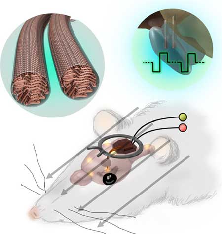
| They are now in " Nature Communication"" On published an article ( " deep brain stimulation and by simultaneously fMRI fibers with the graphene electrode is plotted fully active mode " ), the development of an electrode graphene fibers highly compatible with MRI, this function may DBS Next, the full activation mode is mapped by fMRI. | |
| These complete maps provide a comprehensive picture of how DBS regulates the brain and are very important for revealing the neuromodulatory effects of DBS treatment. | |
| In addition to showing almost no artifacts in various anatomical and functional MRI images, graphene fiber electrodes also show high charge injection capability and stimulation stability, which is very important for DBS electrodes. | |

|
|
| Schematic diagram of a DBS-fMRI study using graphene fiber bipolar microelectrodes. (Reprinted with permission from Springer Nature) | |
| Duan said: "The use of graphene fiber electrodes in DBS enables a complete mapping of activation patterns." "We detected a blood oxygenation level dependent (BOLD) response, which is a signal that the brain is in multiple cortexes and cortex The activation of the lower area. The BOLD reaction in some areas could not be detected with traditional metal electrodes before. Their big artifact. " | |
| The team‘s platform can be used as a powerful tool for the translational study of the neuromodulation and treatment mechanisms of DBS treatment. | |
| Duan pointed out: "With further development and careful safety assessment, graphene fiber electrode technology may even be used as a DBS electrode for patients." Combined with MRI, the clinical effect of DBS treatment can be predicted and the clinical results optimized. " | |
| The researchers pointed out that the DBS-fMRI study using graphene fiber electrode technology is widely applicable to other targets or neural circuits. Therefore, they plan to use graphene fiber electrodes in DBS-fMRI studies of other diseases, especially in the treatment of refractory depression. | |
| The research team hopes that the use of graphene fiber electrodes under DBS on different targets and with different stimulation frequencies and intensities of the whole brain activation pattern mapping can provide important insights for the development of effective DBS treatment of depression. | |
| Although the current study used anesthetized animals for fMRI localization, the difference in physiological status between normal (awake) and fMRI (anaesthesia) may limit the detection of all potential neurological correlations of DBS treatment effects. For future research, the application of functional magnetic resonance imaging in conscious animals will help provide more detailed clues to the circuit mechanism of DBS treatment. | |
|
Duan concluded: "In order to improve the DBS-fMRI study, we first need to resolve the difference between the physiological state of awake and anesthesia." "In addition, another challenge we need to overcome is how to improve the stimulation selectivity and long-term stability of DBS. . We are also committed to improving electrode design and materials. " Source: Nanowerk |
- Previous: 3D printed metal-organ
- Next: A Rising 2D Star: Nove


 Academic Frontier