Bone transplantation is widely used in clinical orthopedic surgery to enhance bone regeneration. Generally, bone tissue engineering generally uses biodegradable scaffolds to fill bone defect sites, and such scaffolds can recruit inorganic components in the body and enhance osteogenic differentiation. In order to further improve the biological activity of the biological scaffold and improve the integration of the scaffold with the surrounding bone tissue, the researchers explored and prepared hydrogels that can autonomously bind inorganic ions and stimulate bone biomineralization, such as chondroitin sulfate (CS)-based hydrogels. Compound hydrogel. CS is a glycosaminoglycan (GAGs) widely present in human bones. Its main role is to coordinate the adhesion of osteoblasts and indirectly participate in the process of bone homeostasis.
In order to explore the role of chondroitin sulfate in bone formation, the research team of Nathaniel S. Hwang, School of Chemistry and Bioengineering, Seoul National University, South Korea, published a paper entitled Chondroitin Sulfate-Based Biomineralizing Surface Hydrogels for Bone Tissue Engineering in ACS Applied Materials & Interfacess. article. As shown in Figure 1, the researchers prepared a PEGDA/MeCS-based composite hydrogel, and through the methacrylylation modification of chondroitin sulfate (Figure 2), the researchers prepared a PEGDA-MeCS composite light-curable bio-ink. It is used to study the biomineralization performance of gel scaffolds.

Figure 1 Schematic diagram of preparation and biomineralization of PEGDA-MeCS composite hydrogel
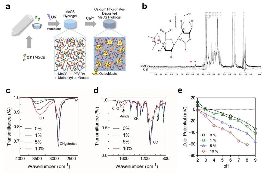
Figure 2 Preparation and physical and chemical properties of PEGDA-MeCS composite hydrogel
Researchers have explored the swelling properties, gel pore size, gel and chondroitinase degradation behavior of the composite gel under different concentrations of MeCS (0%-10%). Experiments have shown that as the concentration of MeCS increases, the swelling rate of the gel gradually increases, and the gel is more prone to degradation (Figure 3).
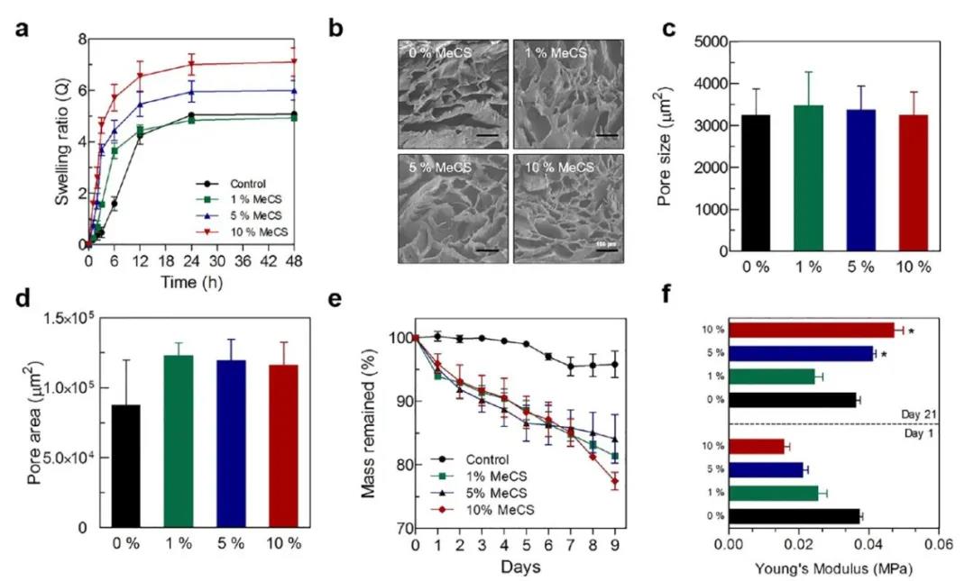
Figure 3 Determination of swelling, pore size, degradation performance and Youngs modulus of PEGDA-MeCS composite hydrogel
Chondroitin sulfate has negatively charged groups and is easy to affinity protein factors and positively charged cations. The researchers immersed the PEGDA-MeCS composite hydrogel in a PBS solution containing calcium ions to explore the affinity of the gel for Ca2+ and phosphate ions. X-ray photoelectron spectroscopy showed that the binding energy of Ca2+ and phosphate ions to the gel increased with the increase of MeCS content. The free calcium ion of the gel was detected by a calcium ion fluorescent probe, and the concentration of free Ca2+ decreased with the increase of MeCS concentration. In contrast, the free phosphate ion in the high-concentration MeCS gel was significantly higher than that in the low-concentration gel group (Figure 4).
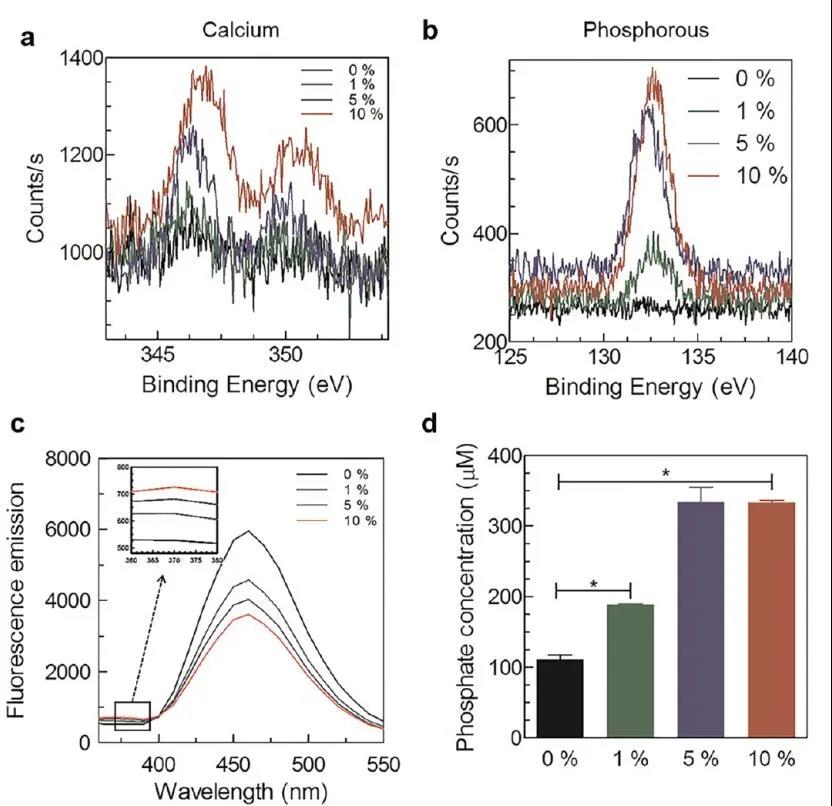
Figure 4 Evaluation of the affinity of PEGDA-MeCS composite hydrogel to calcium ions and phosphate ions
Based on the strong affinity of chondroitin sulfate for calcium ions, the researchers analyzed the white particles formed on the surface of the PEGDA-MeCS composite hydrogel using X-ray energy spectroscopy (EDS). Studies have shown that as the concentration of MeCS increases, the concentration of phosphate and calcium ions on the surface of the hydrogel gradually increases. In order to confirm whether these white particle deposits are calcium phosphate (CaP) derivatives, the researchers compared the blank gel group containing MeCS components. The gel surface of the MeCS gel group with a content of 5% is relatively rough and with high resolution. Transmission electron microscopy (HR-TEM) showed that the CaP formed on the surface of the gel contained a crystalline structure of hydroxyapatite (HAP). In summary, the experiments show that PEGDA-MeCS composite hydrogel can induce the formation of HAP (Figure 5).
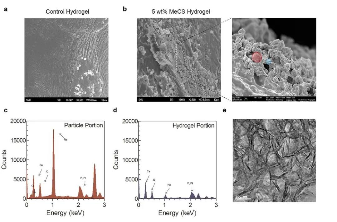
Figure 5 Evaluation of CaP formation performance of PEGDA-MeCS composite hydrogel in vitro
In order to test the biocompatibility of the hydrogel, it is studied to culture human tonsillar mesenchymal stem cells (hTMSC) in PEGDA-MeCS composite hydrogel three-dimensionally. The live-death staining test shows that the gel’s biological activity on cells can be maintained at 84.8%. And the expression of osteogenic-specific genes (osteogenesis-specific transcription factor (RUNX2), osteocalcin (OCN), alkaline phosphatase (ALP) and type I collagen (COL I)) was significantly higher than that of those without MeCS Gel blank group (Figure 6).
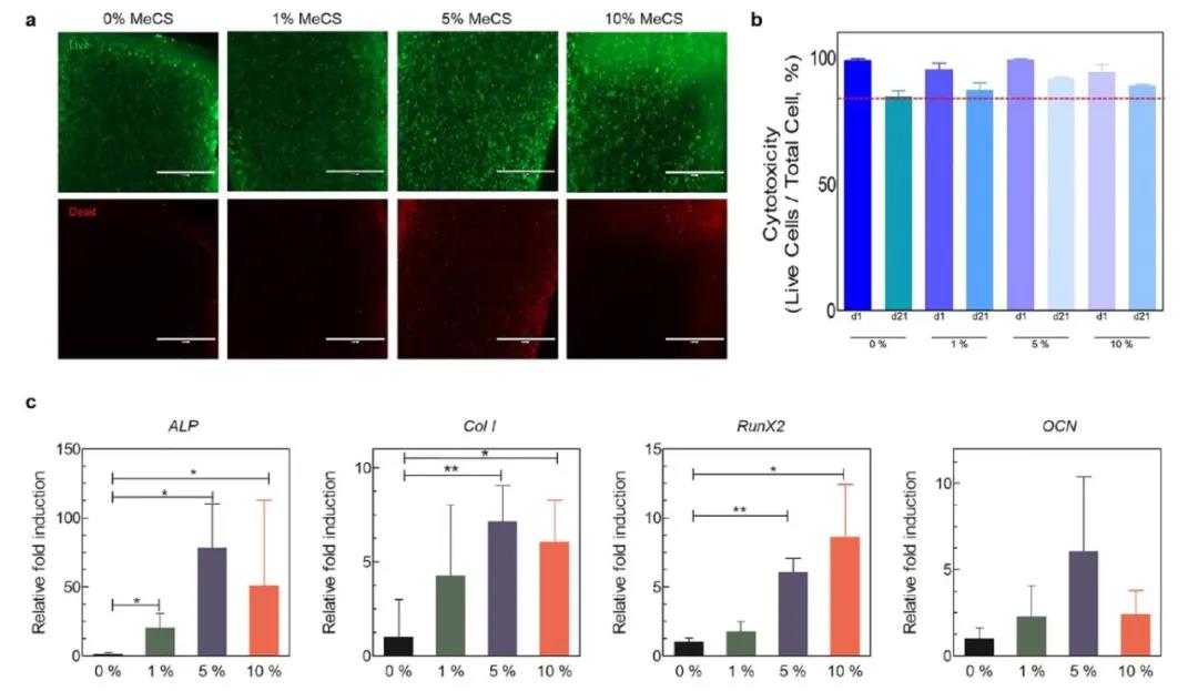
Figure 6 Evaluation of biocompatibility and osteogenic performance of PEGDA-MeCS composite hydrogel in vitro
In order to explore the osteogenic properties of the hydrogel in vivo, the researchers implanted the hTMSC-loaded hydrogel scaffold into BALB/c mice with a 4mm diameter skull defect. After 8 weeks of incubation, Micro-CT scanner test results showed that the volume of new bone in the composite gel group with MeCS content of 5% and 10% was significantly higher than that of the control group. The HE and Masson staining experiments show that the integration of the composite gel with 10% MeCS content with the original bone is better than other groups, and the cell infiltration is better. Immunostaining of type I collagen showed that the collagen in the composite gel with a MeCS content of 10% was evenly distributed on the bone defect, while the collagen in the composite gel group with a MeCS content of 0% and 1% was only distributed on the surface of the gel ( Figure 7).

Figure 7 Evaluation of the osteogenic properties of PEGDA-MeCS composite hydrogel in vivo
In summary, based on the good affinity between chondroitin sulfate and calcium ions and phosphate ions, the researchers prepared a light-cured PEGDA-MeCS composite hydrogel, which provides a good osteogenic micro surroundings. And in vivo and in vitro experiments showed that with the increase of MeCS content, the biomineralization and osteogenic differentiation ability of the composite gel gradually increased. This PEGDA-MeCS-based biomineralization hydrogel provides a potential carrier in clinical applications of bone mineralization and bone regeneration.
Information source: EngineeringForLife
This information is from the Internet for academic exchanges. If there is any infringement, please contact us and delete it immediately