Inflammation refers to the bodys mechanical, biochemical or immune-mediated adaptive response to stimuli. However, overactive inflammatory response is harmful. A prolonged inflammatory process will not only interfere with the repair process of damaged tissues, but also trigger a series of cascade reactions. These cascade reactions and inflammatory cells continue to recruit to the vicinity of the lesion and form a local area. The inflammatory cytokine storm is related, and these two conditions will eventually hinder tissue repair and exacerbate degeneration. Injectable hydrogel microspheres have the advantages of prolonging the half-life of drugs and maintaining the effective concentration of local drugs. They have shown great potential in clinical drug carrier applications, but local hyperactive inflammation in degenerative diseases may affect this treatment effect.
Recently, Professor Chen Liang and Professor Gu Yong from the First Affiliated Hospital of Soochow University and Professor Cui Wenguo from Ruijin Hospital of Shanghai Jiaotong University School of Medicine jointly published a titled "Modulation of Local Overactive Inflammation via Injectable Hydrogel Microspheres" in Nano Letters. In the research paper, the author constructs injectable "peptide-cell-hydrogel" microspheres by covalently coupling APETx2 to methacryloyl gelatin (GelMA) and further loading nucleus pulposus cells, which can both inhibit The local inflammatory cytokine storm also realizes the regulation of the extracellular matrix (ECM) metabolic balance in vitro, and provides an effective way for tissue repair and regeneration under overactive inflammation.
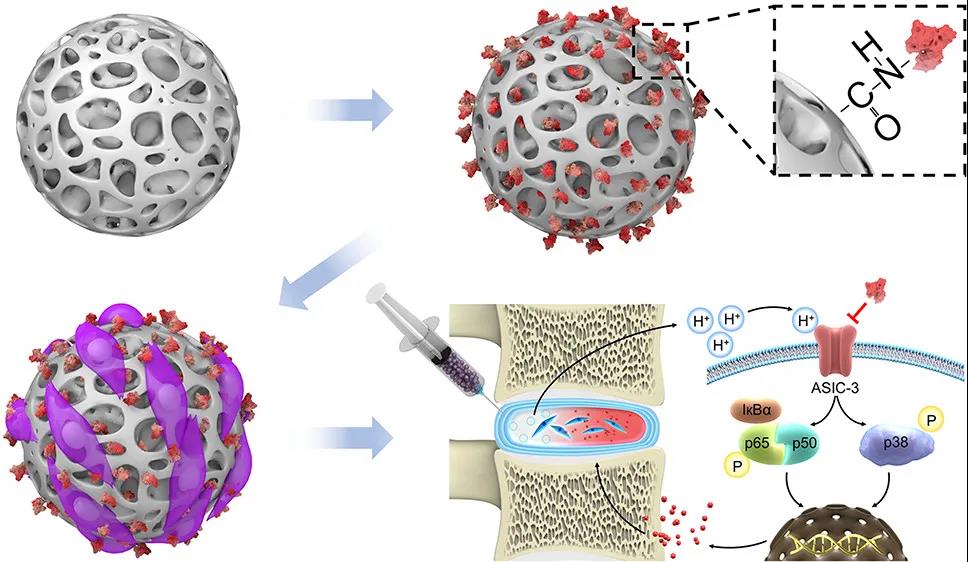
Figure 1 Schematic diagram of the article, coupling APETx2 on GelMA and further loading nucleus pulposus cells to construct injectable "peptide-cell-hydrogel" microspheres, which can inhibit local hyperactive inflammation and maintain ECM stability.
Microfluidic technology is an ideal platform for constructing hydrogel microspheres. It can not only load drugs and cells at the same time, but also is easy to operate. By covalently coupling with the active peptide APETx2 and further carrying nucleus pulposus cells, a "peptide-cell-hydrogel" gel microsphere can be constructed. As shown in Figure 2.: GelMA microspheres (GM) are prepared by a microfluidic synchronous photocrosslinking device, washed and freeze-dried to prepare porous GelMA microspheres; APETx2 conjugated porous GelMA microspheres (GA) are prepared and loaded with marrow Nuclear cells, injected with "peptide-cell-hydrogel" microspheres (GNA) in a rat model of intervertebral disc (IVD) degeneration; "peptide-cell-hydrogel" microspheres regulate the degeneration of IVD in rats The hyperactive inflammation, and promote the proliferation of nucleus pulposus cells and the regeneration of IVD.
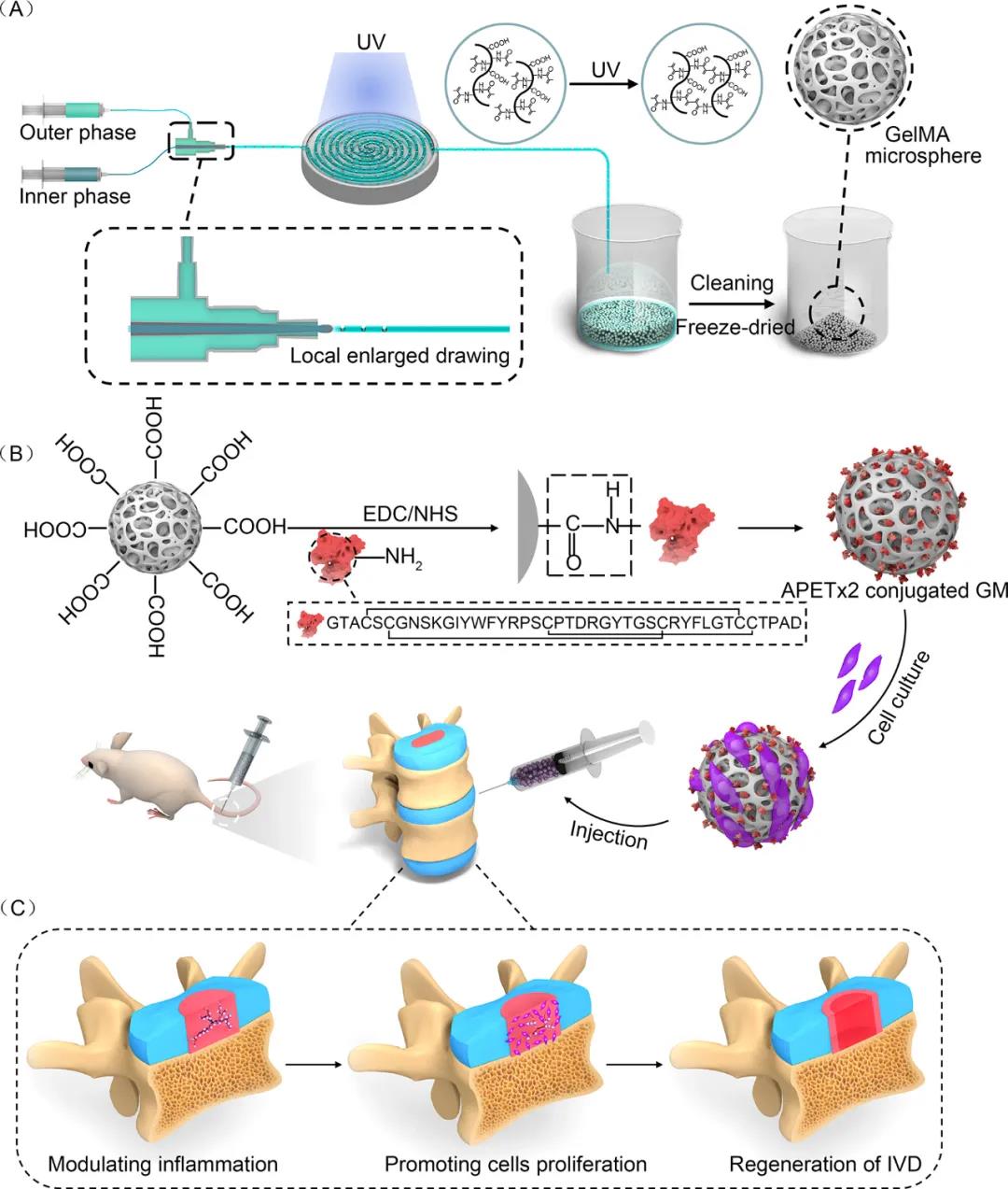
Figure 2 A schematic diagram of injectable "peptide-cell-hydrogel" microspheres to regulate local hyperactive inflammation to promote intervertebral disc regeneration.
The gel microspheres are formed by a water-in-oil method in a microfluidic device, and are further photo-crosslinked and freeze-dried to obtain porous gel microspheres. As shown in video 1, under a microscope, the gel microspheres prepared by the coaxial nozzle shearing in the same direction have good dispersibility and uniform size. The microspheres can smoothly pass through the needle of the micro syringe without damaging the structure.
Video 1 The microsphere passed through the needle of the micro syringe without any damage to the structure.
In order to give nucleus pulposus cells the ability to regulate local hyperactive inflammation, the carboxyl groups on the surface of the microspheres are activated by EDC/NHS and then covalently linked to the amino residues on APETx2 to form APETx2-GelMA microspheres (GA). Next, the XPS analysis and Fourier transform infrared spectroscopy analysis of Fig. 3 showed that the APETx2 peptide was successfully grafted and SEM showed that GA retained the porous structure of GM. Glycolysis is the main form of energy supply to the intervertebral disc, and the gradual accumulation of lactic acid produces an acidic microenvironment in the degenerative intervertebral disc. Therefore, the researchers studied the release behavior of gibberellin under two pH conditions (7.4 and 6.5) through high performance liquid chromatography. The release rate of the peptide under pH 6.5 is higher than that under pH 7.4, but the two There is no significant difference between them.
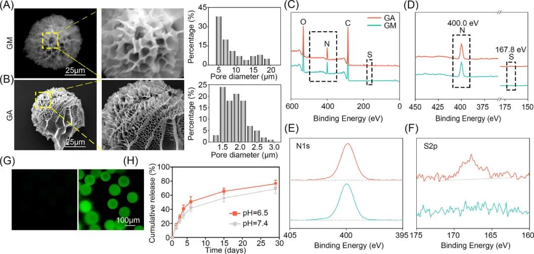
Figure 3 Characterization of injectable "peptide-cell-hydrogel" microspheres (GNA). (A, B) SEM observation of the microscopic morphology differences between GM and GA microspheres; (CF) XPS analysis and Fourier transform infrared spectroscopy of GM and GA microspheres; (GH) fluorescence before and after grafting FITC-BSA Image, and the release curve of GA releasing APETx2 at pH 7.4 and pH 6.5.
In addition, in an acidic environment in vitro, the researchers tested the expression levels of related proteins and genes. As shown in Figure 4, the expression of ASIC-3 protein in the glutamate group was significantly inhibited, and there was no significant difference from the control group. The inflammatory cytokines interleukin-1β, interleukin-6 and tumor necrosis factor-α were also significantly inhibited by APETx2. At the same time, APETx2 down-regulates the expression of MMP-3 and ADAMATS-5, thereby reducing the degradation of extracellular matrix synthesized by NPCs, leading to an increase in the content of extracellular matrix. The high expression of COL2 and aggrecan protein also proves this a little. The above results indicate that APETx2 can inhibit the activation of ASIC-3 and reduce the inflammatory cytokine storm of the intervertebral disc caused by the acidic environment. In addition, APETx2 can reverse the imbalance of extracellular matrix synthesis/catabolism in nucleus pulposus cells in acidic environment, leading to increased extracellular matrix deposition in nucleus pulposus cells and inhibiting intervertebral disc degeneration.
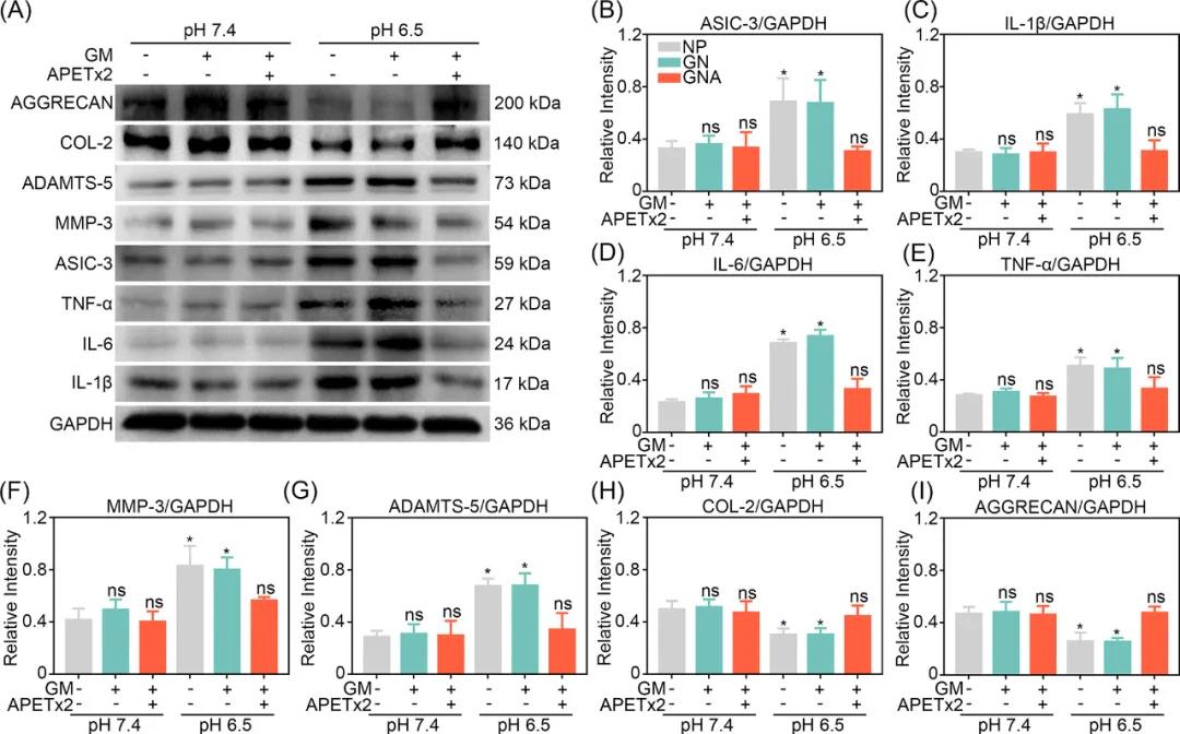
Figure 4 7-day protein and gene expression of nucleus pulposus cells in an acidic inflammatory microenvironment in vitro. (A) Western blot of ASIC-3, inflammatory factors, extracellular matrix degrading enzymes and extracellular matrix proteins. (B-I) RT-PCR semi-quantitative determination of gene expression of ASIC-3, inflammatory factors, and extracellular matrix degrading enzymes.
In order to study the role of GNA in promoting the regeneration of IVD in vivo, the researchers injected GA (GNA) rich in nucleus pulposus cells into a rat model of IVD degeneration. The imaging results in Figure 5 show that adjusting the local inflammatory microenvironment or supplementing the nucleus pulposus cells alone can delay the degeneration of the intervertebral disc, but cannot reverse the degeneration of the intervertebral disc. On the other hand, the "peptide-cell-hydrogel" microspheres not only maintain and restore the high water content in the nucleus pulposus cells, but also promote the regeneration of degenerated intervertebral discs while delaying the degeneration process.

Figure 5 Imaging data of animal experiments. (A) Establishment of rat tail puncture model. (B) Representative x-ray images of rat tail vertebrae at 4th and 8th week. (C-E) Changes in the percentage of DHI in each group in the 4th and 8th weeks. (F) Representative MRI images of rat tail vertebrae. (G) The magnetic resonance grading changes of each group at the 4th and 8th week.
Tissue sections were collected for further analysis at 4 and 8 weeks after surgery. The results are shown in Figure 6. Compared with the normal control group, hematoxylin-eosin and safranin O-Fast Green staining showed the degeneration and structure of the intervertebral disc tissue in the GNA group The damage has improved. In addition, there was a clear and definite boundary between the nucleus pulposus and the annulus fibrosus, and the content of the nucleus pulposus was significantly improved at 8 weeks. However, in the DC, GA and GN groups, the nucleus pulposus of the intervertebral disc gradually ruptured as the disc herniated, and the boundary between the nucleus pulposus and the annulus fibrosus became increasingly blurred. These results are consistent with further research results of immunohistochemical staining. The COL2 content of the GNA group recovered completely at the 8th week, and the histological score showed that the collagen content of the GNA group was significantly higher than the DC, GA and GN groups at the 4th week, but lower than the normal control group. The collagen content of the GNA group recovered at the 8th week, and there was no significant difference from the normal control group, indicating that the "peptide-cell-hydrogel" microspheres can promote the regeneration of degenerative intervertebral discs.
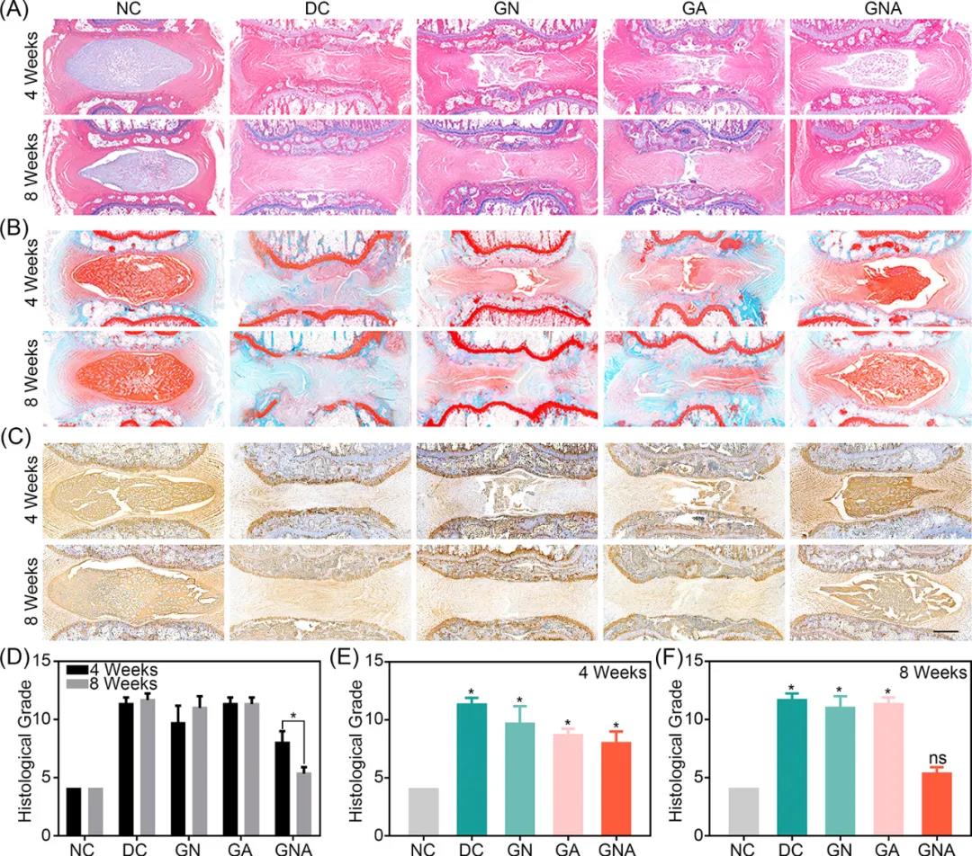
Figure 6 Histological evaluation of animal experiments. (A) Hematoxylin-eosin staining images of rat intervertebral discs at 4 and 8 weeks. (B) Safranin O-fast green staining. (D-F) COL2 immunohistochemical staining. Changes in histological grades in the 4th and 8th weeks of each group.
In summary, this study produced porous "peptide-cell-hydrogel" microspheres covalently grafted with APETx2 and carried nucleus pulposus cells through microfluidic technology, and confirmed this new "peptide-cell" In addition to participating in the tissue regeneration process, "hydrogel" microspheres can also regulate local hyperactive inflammatory responses to maintain the synthesis/catabolism balance of the extracellular matrix in the nucleus pulposus of the intervertebral disc. This injectable "peptide-cell-hydrogel" microsphere provides a new idea for tissue regeneration in the microenvironment of local hyperactive inflammation.