I. Overview of the article.
Phenols are ubiquitous in nature and have attracted wide attention because of their unique physical and chemical properties and a wide range of industrial applications. In recent decades, their accessibility, multifunctional reactivity and relative biocompatibility have promoted the research of phenolic nanotechnology (PEN), especially for biomedical applications, which is the main benefactor of this emergence, which is proved to a large extent by polydopamine and polyphenols. Therefore, it is necessary to summarize the basic mechanism and synthesis strategy of PEN in the most advanced biomedical applications, and provide a timely and comprehensive summary. In this review, we will focus on the principles and strategies involved in PEN, and summarize the use of PEN synthesis toolkits in particle engineering and bottom-up synthesis of nano-hybrid materials. Specifically, we will discuss the attraction between phenols and complementary structural motifs in confined particle systems to synthesize high-quality products with controllable size, shape, composition, surface chemistry and functions. In addition, many applications of phenolic in biosensor, biological imaging and disease treatment will also be emphasized. The purpose of this review is to provide guidelines for new scientists in this field and to serve as the latest compilation of achievements in this field, as well as expert views on the use of PEN in transformation research.
Second, guided reading of picture and text.

Figure 1.
Development of PEN for biomedical applications. From left to right: representative natural resources of phenols, chemical structure of representative phenolic molecules, some typical PEN-mediated particle diagrams, and potential applications in biomedical fields, including biomedical nanointeraction research, biosensors, imaging and disease therapy, from single molecular and cellular levels to animal and clinical trials.

Figure 2.
All-atomic MD simulation of pBDT (blue) and catechin (red) assembly in water at 0,0.5,4.0 and 10 ns. The enlarged interface shows the aromatic accumulation of BDT and CAT and the hydrogen bond (red line) between CAT and water; carbon, oxygen, hydrogen and sulfur are black, red, white and yellow, respectively.

Figure 3.
Phenolic particles co-assembled with metal ions and small molecular drugs. (a) the chemical structure of the structural units (Pt-OH, PEG b-PPOH) and the schematic diagram of phenolic particles prepared by copolymerization. (B) TEM images of phenolic particles prepared. (an and b) reproduced with permission. Copyright 2020 Wileymuri VCH. (C) schematic diagram of the synthesis of carrier-free nano-bomb (DNPs/N@PDA) with PDA coating and NIR response. (d) TEM images of DOX nanoparticles (DNPs), DNPs coated by PDA (DNPs@PDA) and DNPs@PDA loaded with NH4HCO3 (DNPs/N@PDA). (cMurf) reproduced with permission. Copyright 2018 Wileymuri VCH.
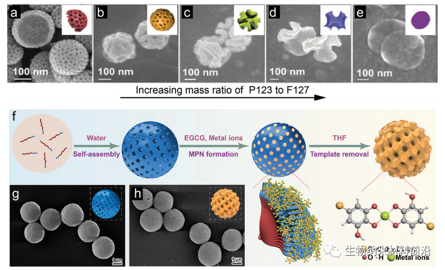
Figure 4.
Soft templates guide the synthesis of mesoporous phenols. The SEM images of PDA particles prepared by different mass ratios of P123 and F127 are as follows: (B) 0: 1; (c) 1: 15; (d) 1: 3; (e) 1: 1; (f) 5: 3. Illustration: schematic diagram of mesophase transition of PDA particles at different P123/F127 mass ratios. (F) A schematic diagram of the synthesis of mesoporous MPN particles using polymer cube (PC) as a template. After removing the template, (g) PCs and (h) SEM images of mesoporous MPN coating PCs.
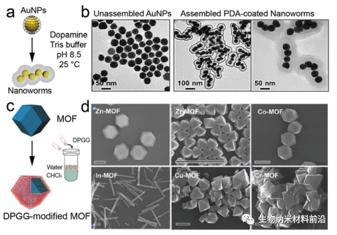
Figure 5.
The viscous phenolic coating was deposited in situ on the preformed nanomaterials. (a) A schematic diagram of the assembly of dopamine-mediated Au nanoparticles (AuNPs) into PDA-coated nano-worms. (B) TEM images of unassembled AuNPs and assembled nano-worms. (C) schematic diagram of surface functionalization of MOF particles by phase transfer reaction. (d) SEM images of DPGG modified MOF particles of different metal ions. Scale: 1 mm.
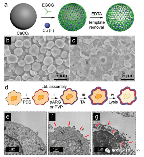
Figure 6.
The hollow phenolic particles were removed by phenolic coating and template. (a) schematic diagram of EGCG-Cu (II) capsule synthesis. (B) SEM images of EGCG-Cu (II) @ CaCO3 nanoparticles and (c) EGCG-Cu (II) capsules. (d) preparation of alternating layers of polyelectrolyte dextran sulfate (PDS), poly-L-arginine (pARG) or poly-L-arginine (pARG) with electrostatic interaction (step i-iii) schematic diagram of the LbL-coated biological hybrid cancer cell template capsule (N-vinylpyrrolidone) (PVP) and TA by hydrogen bond, and then cleavage the cell (iv) after hypotonic treatment. (e) TEM images of uncoated cells, (f) double-layer PVP/TA-coated cells and (g) double-layer TA/PVP-coated cells. The red arrow indicates the LbL coating.

Figure 7.
Study on cellular absorption of phenolic nanoparticles. (a) the uptake, transport and accumulation of PDA nanoparticles in cells. (B) TEM images, (c) enlarged TEM images and (d) confocal laser scanning microscope images of cells incubated with PDA nanoparticles for 3 days. (e) the scheme of inner body escape of naked nanoparticles and MPN-coated nanoparticles and fluorescence microscope images.
III. Summary of the full text.
Phenolic compounds show great potential in nanotechnology, especially in the design of multi-functional nanosystems. This work is based on an excellent summary of published phenolic hydrogels or integral coatings. Here, we summarize the development of multifunctional nanosystems (for example, core-shell nanoparticles, mesoporous nanoparticles, Janus nanoparticles, etc.), which are composed of phenolic compounds through covalent bonds or with various structural units (e.g., small molecules, DNA, peptides, proteins, metals, semiconductors, etc.). In addition to the biocompatibility of phenolic resins, the unique physical and chemical properties of these nanosystems can further determine their functions in biomedicine. Extensive achievements have been made in the research community, but a better understanding of complex material-material interactions (i.e. between phenolic resins and other different building blocks) and their complex bio-nano interactions (for example, interactions between PEN nanoparticles and different biological environments) is important. Although many researchers have developed and applied phenolic materials in the past few decades, significant progress has been made in promising biomedical applications. We envisage that the joint efforts of nanotechnology, organic chemistry and medical researchers will provide higher controllability for phenolic nanoparticles, thus expanding their applications in biomedicine and other fields.
This information is from the Internet for academic exchange only. if there is any infringement, please contact us to delete it immediately.