已传文件:photo/1793410855.png

The work entitled "Sea Urchin-Inspired Immuno-Instructive Ionic Flow Drives MSCs-Macrophage Communication to Promote Bone Regeneration" has been published in Advanced Materials.
Bone tissue regeneration is a complex physiological process that relies on the spatiotemporal coordination of immune cells and stem cells. Traditional studies have mainly elucidated the mechanisms by which materials promote bone formation by first modulating macrophages and subsequently stimulating osteogenic differentiation of stem cells. However, these studies have largely overlooked the active regulation of immune cells by stem cells. Therefore, investigating the capacity of materials to recruit stem cells and concurrently modulate innate immune infiltration, as well as establishing responsive mechanisms of the tissue microenvironment, is of great significance for the development of next-generation tissue regeneration materials. In this study, a sea urchin-inspired immunoinstructive ion flux (SUIF) platform with a radial mesoporous structure was introduced. This platform generates a "fast-slow biphasic" ion release profile via the dissociation/dissolution kinetics of B─O bonds and sustained Sr²⁺ release, thereby constructing a dynamic alkaline ion microenvironment. Within this microenvironment, 5B5Sr-SUIF recruits mesenchymal stem cells (MSCs) and stimulates MSCs to upregulate the expression of miR-466m-5p, which blocks the nuclear translocation of NF-κB in macrophages. This facilitates immunoregulatory crosstalk between MSCs and macrophages, offering new material design principles and mechanistic insights for the precise regeneration of complex bone defects.
Quick Look at the Literature
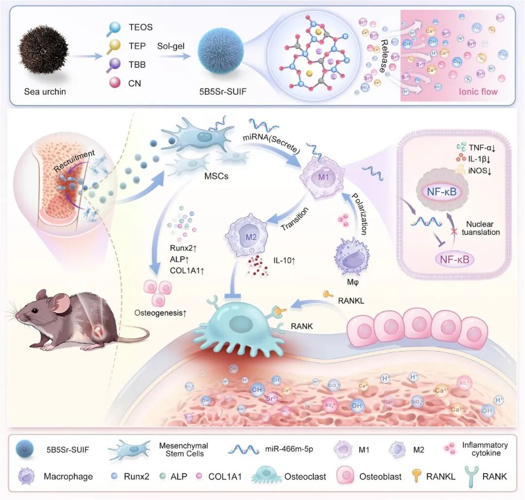
Figure 1
Schematic diagram of 5B5Sr-SUIF synthesis and its potential mechanism for promoting communication between mesenchymal stem cells and macrophages through multifunctional ion flow, thereby enhancing bone regeneration.
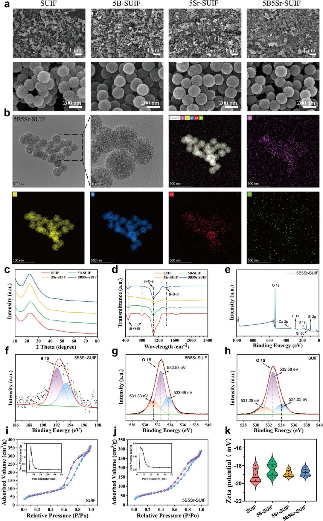
Figure 2
Morphological and physicochemical characterizations of SUIF-based nanoparticles. (a) SEM images of SUIF, 5B-SUIF, 5Sr-SUIF and 5B5Sr-SUIF. (b) TEM image and the corresponding elemental mapping of 5B5Sr-SUIF. (c) XRD patterns of SUIF, 5B-SUIF, 5Sr-SUIF and 5B5Sr-SUIF. (d) FT-IR spectra of SUIF, 5B-SUIF, 5Sr-SUIF and 5B5Sr-SUIF. (e) Full XPS survey spectrum of 5B5Sr-SUIF. (f) High-resolution B 1s XPS spectrum of 5B5Sr-SUIF. (g) High-resolution O 1s XPS spectrum of 5B5Sr-SUIF. (h) High-resolution O 1s XPS spectrum of SUIF. (i) N₂ adsorption-desorption isotherm of SUIF. (j) N₂ adsorption-desorption isotherm of 5B5Sr-SUIF. (k) Zeta potential curves of SUIF, 5B-SUIF, 5Sr-SUIF and 5B5Sr-SUIF.
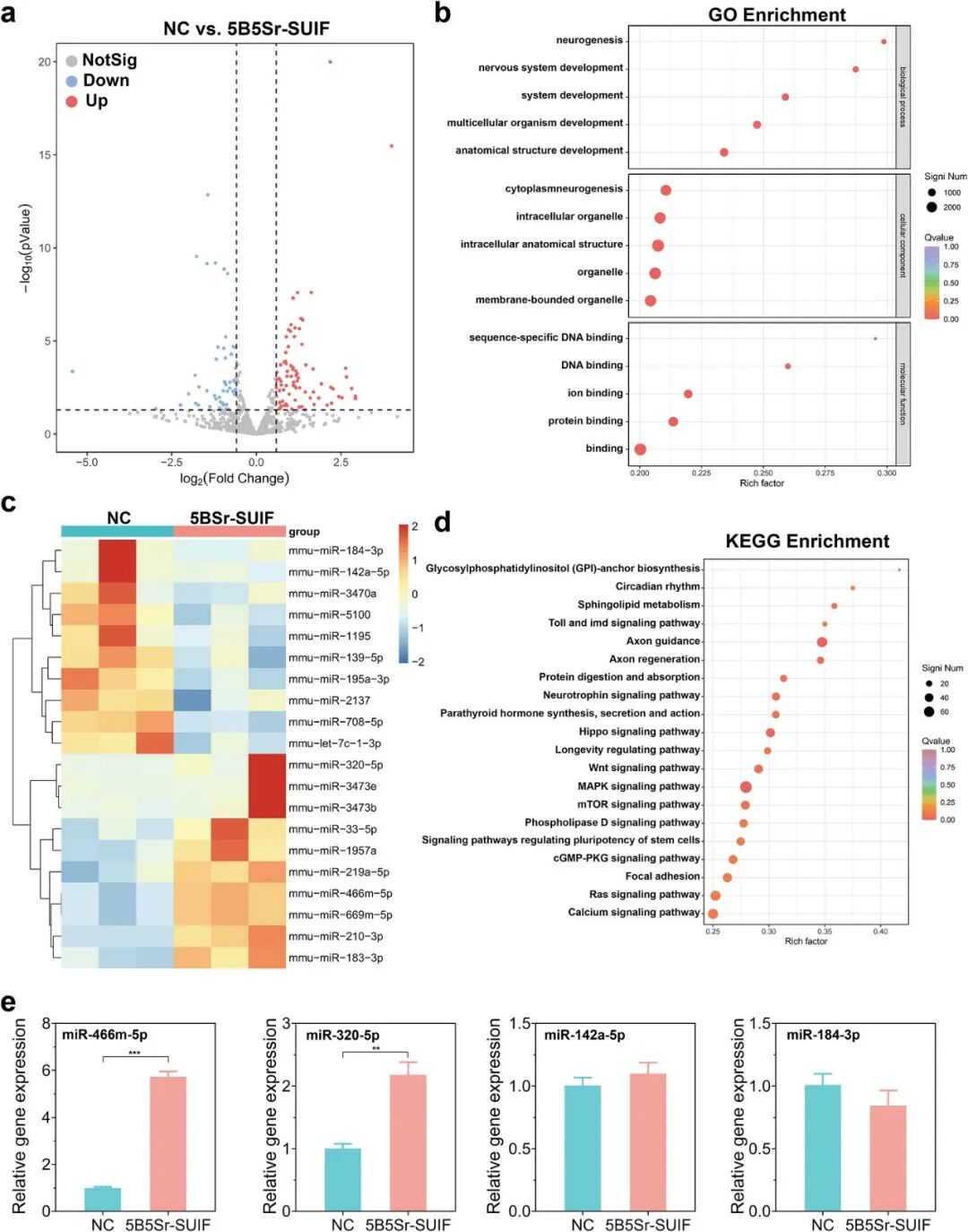
Figure 3
MicroRNA (miRNA) sequencing analysis of the control group (NC group) and 5B5Sr-SUIF group. (a) Volcano plot of differentially expressed microRNAs. Red dots indicate significantly upregulated microRNAs, blue dots indicate significantly downregulated microRNAs, and gray dots represent microRNAs with no significant difference in expression. (b) GO enrichment analysis of differentially expressed microRNAs; the vertical axis denotes the classification of GO terms. (c) Hierarchical clustering heatmap of differentially expressed microRNAs. (d) KEGG bubble diagram showing the top 20 signaling pathways in terms of enrichment degree. (e) Quantitative real-time PCR (qPCR) was used to verify the expression levels of miR-466m-5p, miR-320-5p, miR-142a-5p and miR-184-3p.
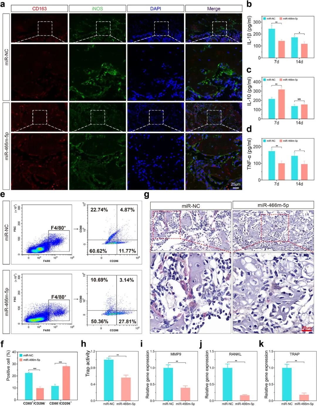
Figure 4
In vivo effects of miR-466m-5p. (a) Immunofluorescence staining of iNOS and CD163 in the tissues around defects after local injection of miR-466m-5p mimic or miR-NC for 2 weeks. ELISA quantification of inflammation-related cytokines IL-1β (b), IL-10 (c) and TNF-α (d) in the defect regions. (e) Flow cytometry analysis of macrophage surface markers CD80 and CD206 in tissue-derived cells. (f) Quantitative analysis of flow cytometry data. (g) TRAP staining of tissue sections from the miR-466m-5p group and miR-NC group. (h) Quantitative evaluation of TRAP activity in tissues of the miR-466m-5p group and miR-NC group. (i–k) qPCR analysis of osteoclast-related genes MMP9 (i), RANKL (j) and TRAP (k) in each group.
Reference Information
Advanced Materials (IF 26.8)
Pub Date: 2026-05-07
DOI: 10.1002/adma.73289
We specialize in custom nano-medical functional materials. We conduct targeted development to tailor structure, particle size and surface modification for your experiments. Full characterization, iterative optimization and technical support are available for targeted delivery, bioimaging and nanomedicine studies. Send your evaluation plans to us.
Engineer Zhang: 17715390137
Engineer Chen: 18914047343