已传文件:photo/1793412295.png
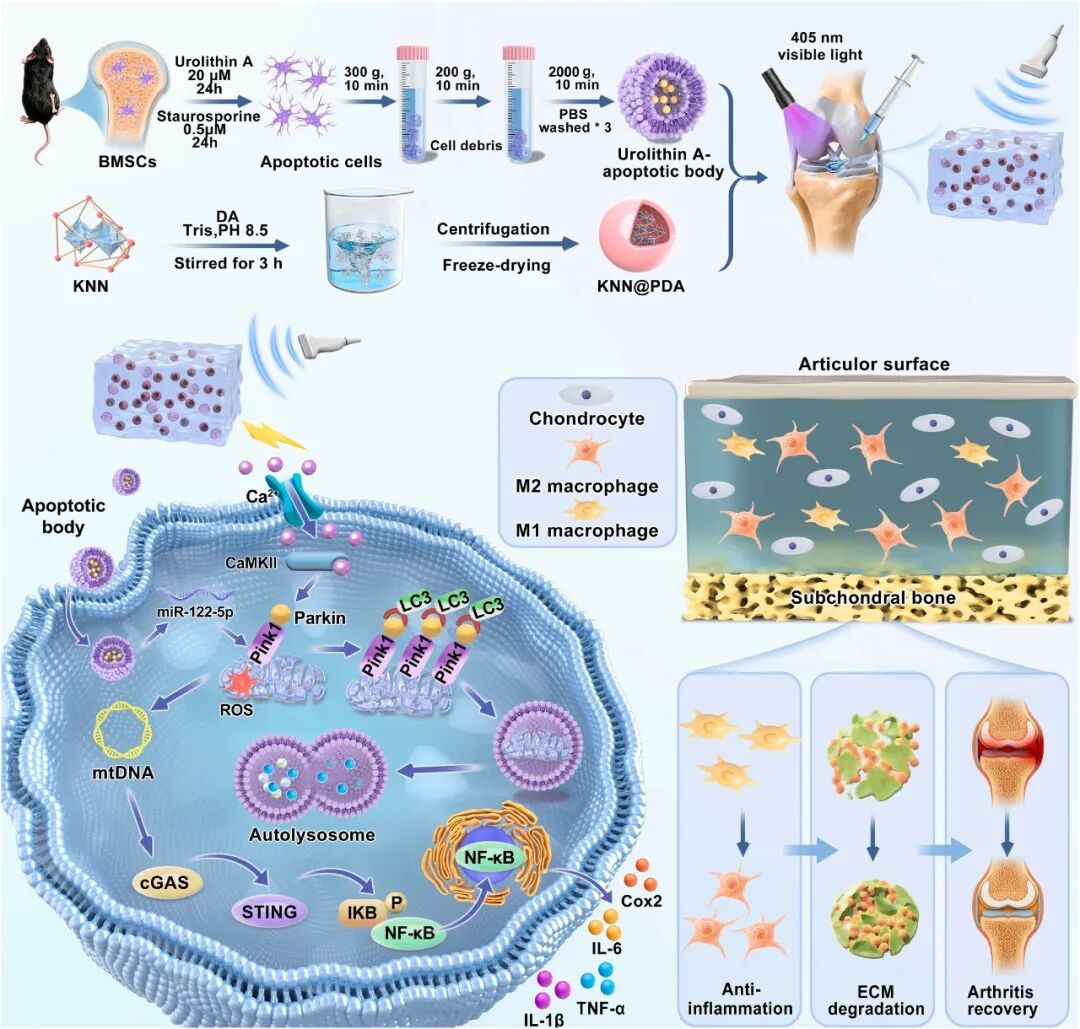
 Osteoarthritis is a degenerative whole-joint disease that involves mitochondrial dysfunction, an imbalanced inflammatory microenvironment, extracellular matrix degradation, and abnormal remodeling of subchondral bone, making it hard for a single biological intervention or physical stimulus to coordinate everything. The study developed an injectable ultrasound-responsive hydrogel GM@KP-ABs, loading polydopamine-modified potassium sodium niobate nanoparticles and urolithin A-engineered apoptotic bodies into a methacrylated gelatin matrix. KNN@PDA can convert low-intensity ultrasound into piezoelectric stimulation, which is linked to enhanced PINK1/Parkin-related mitophagy, while UA-ABs provide mitochondrial regulatory vesicle signaling. The combined treatment is associated with better damaged mitochondria clearance, improved mitochondrial function indicators, weakened mtDNA-related cGAS-STING-NF-κB signaling, M2-like macrophage polarization, reduced inflammation, and improved ECM metabolism balance, and it also alleviates cartilage degeneration and abnormal subchondral bone remodeling in the DMM model.
01
Research Background
 The pathological processes of osteoarthritis are intricately intertwined. Mitochondrial damage promotes ROS accumulation, mtDNA release, and the activation of inflammatory pathways including cGAS‑STING‑NF‑κB, while impaired mitophagy sustains injury and inflammation. Current symptomatic treatments are unable to halt disease progression, and strategies integrating biological and physical cues targeting mitochondrial quality control are therefore required. Apoptotic bodies carry proteins, lipids and nucleic acids derived from parental cells, and pretreatment with urolithin A can endow them with mitochondrial regulatory cues. Nevertheless, free vesicles exhibit limited intra‑articular retention and functional durability. Piezoelectric materials can generate localized electrical cues, and ultrasound‑responsive piezoelectric hydrogels enable remote activation. However, most existing systems prioritize physical stimulation with insufficient biochemical regulation. Integration of engineered apoptotic bodies and piezoelectric hydrogels holds great potential for constructing a bioelectricity‑vesicle coupled platform.
02 Main Content
This study designs an injectable hydrogel integrating piezoelectric conversion and vesicle signaling. KNN@PDA serves as an ultrasound-responsive electroactive transduction unit, while UA-ABs act as vesicle units for mitochondrial and immunomodulation, both dispersed within the GelMA network. Upon ultrasound activation, the hydrogel generates piezoelectric electrical cues and accelerates the release of vesicle-associated components; UA-ABs deliver cargos such as miR-122-5p. Together, they exert therapeutic effects via PINK1/Parkin-dependent mitophagy, clearance of damaged mitochondria, the mtDNA-cGAS-STING-NF-κB axis, macrophage polarization, inflammation regulation and ECM metabolism. The chondroprotective and subchondral bone-protective effects are evaluated in a DMM-induced osteoarthritis model.
03 Study Design
KNN@PDA and UA-ABs are prepared and characterized, followed by the fabrication of GM@KP-ABs. Its injectability, porous architecture, swelling behavior, compressive mechanical properties, rheology, degradation profile, XRD patterns, PFM piezoelectric response, ultrasound-triggered voltage output, vesicle-associated protein release, intra-articular retention and biocompatibility are assessed. In vitro, chondrocytes are stimulated with IL-1β, and a panel of assays including RNA sequencing, membrane potential and Ca²⁺ imaging, JC-1 staining, MitoSOX staining, autophagic flux reporting, mitochondria-lysosome colocalization, western blotting and immunofluorescence are performed. Macrophage polarization and inflammatory cytokines are also evaluated. Mechanistic validations are conducted via PINK1/Parkin intervention, as well as gain- and loss-of-function assays of Mdivi-1 and miR-122-5p. For in vivo experiments, the DMM OA model is adopted with intra-articular injection combined with ultrasound treatment. Histology, OARSI scoring, immunohistochemistry/immunofluorescence, micro-CT and systemic biosafety are examined.
04 Results
GM@KP-ABs features injectability, interconnected porous structure, soft mechanical performance and sustained-release behavior. It maintains the crystalline phase of KNN and local piezoelectric responsiveness, produces voltage output under ultrasound and facilitates accelerated release of vesicle-related proteins, with prolonged intra-articular retention. In vitro, the ultrasound-activated group preserves mitochondrial membrane potential, reduces mitochondrial superoxide, enhances autophagic flux and mitochondria-lysosome colocalization, upregulates PINK1, Parkin and LC3B-II, and downregulates p62. It suppresses cGAS, STING and p65 phosphorylation, diminishes inflammatory mediators, promotes the M2-like macrophage phenotype, restores Collagen II and Aggrecan, and decreases MMP13. miR-122-5p participates in UA-ABs-mediated mitophagy and inflammatory inhibition; Parkin knockdown abrogates the protective effects and reactivates inflammation. In vivo, the hydrogel alleviates cartilage degeneration, downregulates ADAMTS5, rescues the expression of Aggrecan/SOX9/Collagen II, reduces MMP13, and improves subchondral bone parameters, with no obvious abnormalities observed in major organs.
05 Outlook
This study demonstrates that piezoelectric electrical cues and engineered vesicle signals can be coupled within an injectable platform to remodel the osteoarthritic microenvironment through mitochondrial quality control. Future work will disentangle the individual contributions of ultrasound per se, piezoelectric activation and ultrasound-accelerated vesicle release, and quantify local electric fields and ultrasound dosages. Mitochondria-specific autophagic flux reporters will be used for direct dynamic monitoring. The direct targets of miR-122-5p and TGFBR1 regulation will be clarified. Long-term fate and chronic safety of KNN@PDA will be tracked. Further validations will be carried out in primary cells, cartilage/osteochondral explants and OA models with distinct phenotypes. Ultrasound parameters, material distribution and batch-to-batch consistency will also be optimized.
Source Information
Bioactive Materials
Pub Date: 2026-09-22
DOI:10.1016/j.bioactmat.2026.09.018
Zhe Zhang, Xiangheng Guan, Yunhao Zhai, Hongxin Lin, Anna Ying, Jiahao Lin, Chenggong Zhu, Binquan Huang, Nuo Chen, Kaihang Zhang, Xuanyong Liu, Jiahao Ying
We specialize in custom nano-medical functional materials. We conduct targeted development to tailor structure, particle size and surface modification for your experiments. Full characterization, iterative optimization and technical support are available for targeted delivery, bioimaging and nanomedicine studies. Send your evaluation plans to us.
Engineer Zhang: 17715390137
Engineer Chen: 18914047343