
hotline:
17715390137
Tel/Wechat:
18101240246 (Technology)
0512-68565571
Email:mxenes@163.com (Sales Engineer)bkxc.bonnie@gmail.com
Scan the code to follow or search the official account on WeChat:
2D Materials Fronrier After paying attention,
click on the lower right corner to contact us,
Enter enterprise WeChat.
Professional Services Online

Exocrine proteins derived from tumors have become important biomarkers in tumor diagnosis, but a large number of exosomes derived from normal cells hinder the accuracy of quantification. Professor Yang Chaoyong of Xiamen University established an adventitia in situ ligation system with double-target specific nucleic acid aptamer recognition activation, and combined with microdrop digital PCR (ddPCR) technique to quantitatively detect tumor-derived exosome PD-L1 (Exo-PD-L1). The authors made use of the high affinity, good recognition selectivity and high sensitivity of ddPCR of aptamers to achieve high sensitivity and selectivity to tumor-derived Exo-PD-L1. This strategy can distinguish between cancer patients and healthy blood donors and has been identified as a more reliable tumor diagnostic marker than total Exo-PD-L1 for the first time. In short, this tracer strategy has great potential in transforming exosomes into reliable clinical indicators and exploring their biological functions.

Fig. 1. Characteristics of A375 exosome and verification of two affinity probes. A) the schematic diagram of the connection of the two affinity probes. The exocrine A375 was characterized by B) transmission electron microscope and C) nanoparticle tracking analysis. D) the binding of MJ5C-L and SYL3C-L to A375 exosomes was analyzed by flow cytometry.
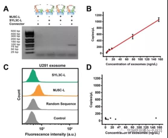
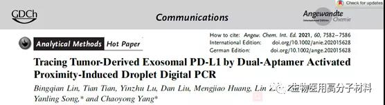
Fig. 2. PLA verification of PD-L1 and EpCAM double positive A375 exocrine and PD-L1 single positive U251 exocrine. A) the gel electrophoresis analysis of the amplified PLA ligated products. B) ddPCR results of A375 exocrine. C) MJ5C-L and SYL3C-L probes against U251 exosomes were analyzed by flow cytometry. D) ddPCR results of U251 exocrine.
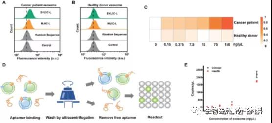
Figure 3. The effects of MJ5C-L and SYL3C-L on exocrine bodies of A) cancer patients and B) healthy donors were analyzed by flow cytometry. C) ddPCR thermograms of exocrine at different concentrations in cancer patients and healthy blood donors. D) the flow chart of ddPCR ultracentrifugation washing quantitative Exo-PD-L1 based on aptamer. E) quantitative results of aptamer-based immune-ddPCR in cancer patients and healthy blood donors.
Figure 4. (A) used ddPCR to detect Exo-PD-L1 in peripheral blood of 33 cancer patients and 12 healthy blood donors. The corresponding ROC curve of B) (A).
This information is from the Internet for academic exchange only. if there is any infringement, please contact us to delete it immediately.

| Reminder: Beijing Beike New Material Technology Co., Ltd. supplies products only for scientific research, not for humans |
| All rights reserved © 2019 beijing beike new material Technology Co., Ltd 京ICP备16054715-2号 |