Extracellular vesicles EV, as nano/micron-sized carriers, have shown great promise in drug delivery and bioimaging. At present, a large amount of research work has explored the unique properties of EVs. Their physicochemical properties, biological characteristics and mechanical properties make them unique carriers and have special pharmacokinetics and circulation in drug delivery. Metabolism and biodistribution patterns. This article first analyzes the pros and cons of EV as a delivery platform. Second, compared with engineered nanoparticle delivery systems such as biocompatible diblock copolymers, a reasonable design scheme for engineered EVs especially exosomes is proposed. Finally, it compares different drug loading strategies for EVs, and provides a reference for how to construct clinically available and efficient nano/microcarriers to achieve satisfactory medical goals.

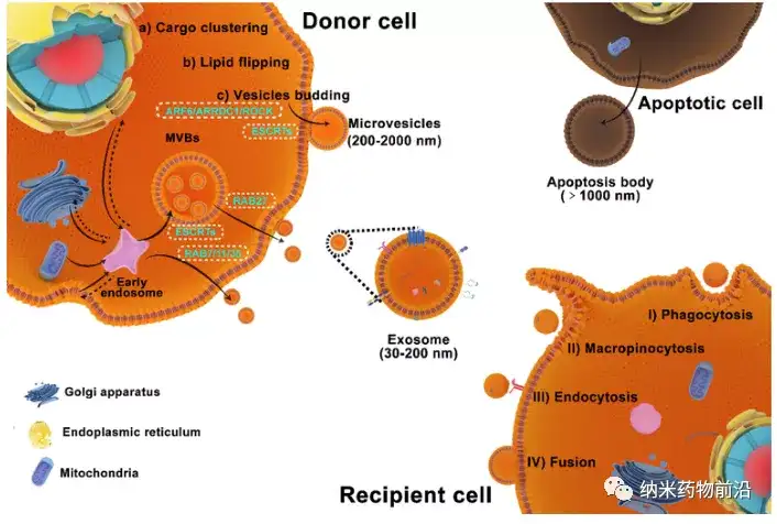
Figure 1 Biological origin and cellular uptake of extracellular vesicles
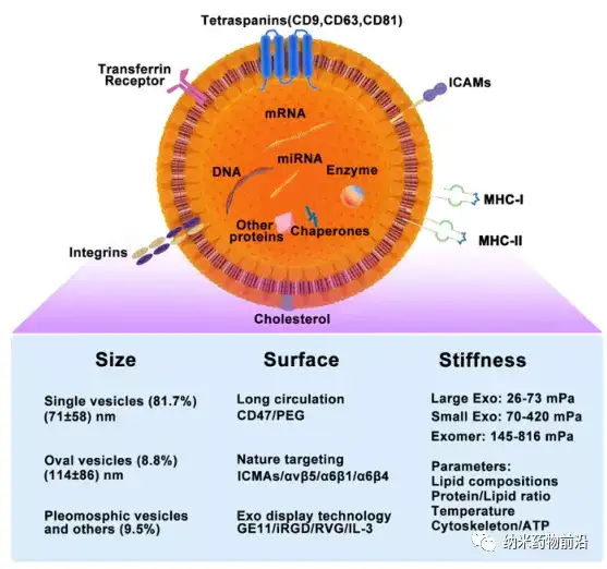
Figure 2 The structure, content, and biomechanical properties of exosomes. Exosomes have a lipid bilayer membrane structure, and their membrane surface highly expresses four-span membrane proteins CD9, CD81 and CD63 , abundant four-span membrane protein related proteins ICMA, integrins, etc. Large amounts of DNA, different types of RNA, enzymes and other functional proteins are encapsulated in exosomes.
A summary of the schematic diagram showing the principle of measuring the mechanical properties of EVs with atomic force microscopy AFM and the atomic force microscope images of red blood cell EVs. The expression of aquaporin-1 AQP1 on EVs can increase its deformability, and the stiffness of AQP1-deficient EVs is significantly higher than that of control EVs. In addition, ultracentrifugation and ultrasonic treatment of the EV can change its rigidity to a certain extent. Surface modification for example, functionalization with polymers or lipids will also change the mechanical properties of EVs. Similar to synthetic particles, changing the polymer type, length, density/coverage or changing the composition/phase behavior of phospholipids will change one. A series of physical and chemical characteristics determined by mechanical properties, including particle size, shape, chemical composition and surface ligands, thereby changing the delivery efficiency of EVs.
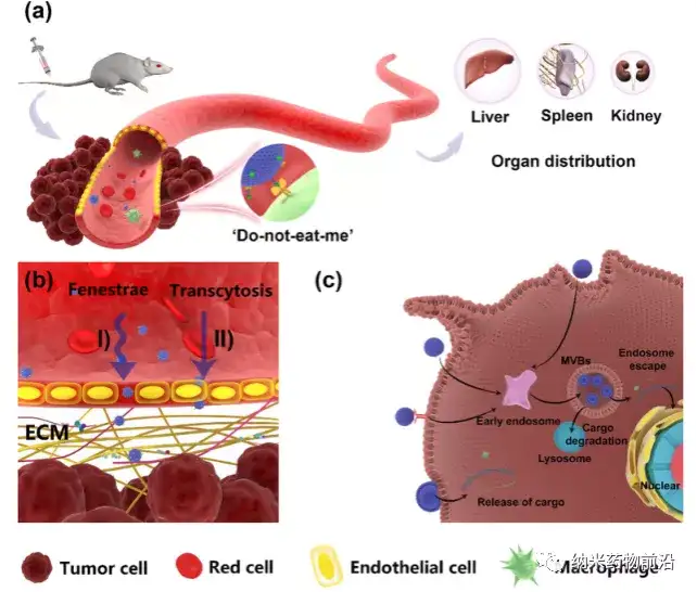
The biological circulation of EV in the process of tumor drug delivery. (a) EVs are mainly accumulated in the liver, spleen and kidney after injected into mice through the tail vein. The expression of CD47 on its surface helps to resist the phagocytosis of macrophages. (b) EVs penetrate vascular endothelial cells in two ways: (1) The deformability of EVs allows them to pass through the interendothelial fenestration passive extravasation; (2) Transcellular transport is the active uptake of EVs by endothelial cells, and then through exocytosis Action to release cargo (c) EVs can directly release cargo after being taken up by the cells by EVs that have passed through membrane fusion.
references
[1] P. Fu, J. Zhang, H. Li, M. Mak, W. Xu, Z. Tao, Extracellular vesicles as delivery systems at nano-/micro-scale, Adv Drug Deliv Rev, (2021) 113910.
This information is sourced from the Internet for academic exchanges. If there is any infringement, please contact us to delete it immediately


