
hotline:
17715390137
Tel/Wechat:
18101240246 (Technology)
0512-68565571
Email:mxenes@163.com (Sales Engineer)bkxc.bonnie@gmail.com
Scan the code to follow or search the official account on WeChat:
2D Materials Fronrier After paying attention,
click on the lower right corner to contact us,
Enter enterprise WeChat.
Professional Services Online

【introduction】
Since the advent of additive manufacturing (commonly known as 3D printing) technology, this technology has revolutionized the field of biomanufacturing and promoted many key advancements in tissue engineering and regenerative medicine. Specifically, compared with the traditional 2D technology, there have been more documents to prove that the rigid monolayer culture system can not recover the complexity inherent in the natural environment. Therefore, the growth under this 2D condition It is difficult for cells to reflect in vivo function, phenotype, morphology, and differentiation potential, and thus is highly affected by what is called extracellular matrix (ECM). Therefore, the 3D cell culture system has gained wide appeal in the fields of tissue engineering and regenerative medicine. At the same time, in order to correctly simulate the 3D ECM environment, a manufacturing method capable of accurately controlling the mechanical, physical and viscoelastic properties of the material in the 3D space is needed. Progress from the latest 3D printing technology shows that they are expected to meet these requirements. The level of control provided by 3D printers has enabled many significant advances in the production of physiologically relevant biomimetic tissue and organ substitutes, such as drug testing, elucidation of biological mechanisms, disease models, translation of medical and surgical implants, etc. In fact, since Dr. Charles Hull first introduced stereolithography (SLA) to the world, many 3D printing technologies have also been developed in a short time. However, the corresponding 3D printing materials have not been developed, which is also a bottleneck restricting the development of this field for some time. In the last ten years, researchers have gradually realized the importance of developing 3D printing materials to maximize the true potential of 3D printing technology.
Recently, the University of California, San Diego (UCSD) Nano Engineering Chen Shao Chen Jiaoshou (Shaochen Chen) (corresponding author) reviewed the development of biological material suitable for light-based 3D printing technology, and its applications focus on biological aspects of print. First, the author introduces the basic principles and mechanisms of photopolymerization in photocurable biomaterials, and summarizes commonly used photoinhibition and photolabile chemicals to control polymerization kinetics. Subsequently, the current literature on photopolymerized natural, synthetic, and composite biomaterials used in light-based 3D printing and their applications in tissue engineering and regenerative medicine are discussed. Finally, the author reviews the recent progress and evolution of optical-based 3D printing technology from serial to flat to volume construction, and discusses strategies to improve print resolution and quality control to standardize future print optimization methods. Overall, expanding and developing new types of photocurable biomaterials will help promote and expand the use of photo-based 3D printing technologies. Related research results were published in Chem. Rev. with the title of " Photopolymerizable Biomaterials and Light-Based 3D Printing Strategies for Biomedical Applications " .
【Graphic introduction】
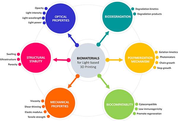
Figure 1. Overview of biomaterial selection criteria for optical-based 3D printing technology in tissue engineering and regenerative medicine
Figure 2. Radical-induced thiol-ene chemical reaction
Figure 3. Effect of olefin group selection on thiol-ene reaction kinetics
(A) The theoretical calculation of the thiol-ene reaction kinetics depends on the reactivity of the selected olefin group;
(B) Decrease in olefin group reactivity based on theoretical kinetic model.
Figure 4. Hydrogel network depending on different cross-linking mechanisms and resulting unevenness
(A) Free radical chain growth polymerization of monomer and cross-linking agent leads to spatial non-uniformity in the network structure;
(B) The functional groups of the polymer chain form a network through crosslinking in a semi-static solution, resulting in local unevenness
(C) Aggregate to form a basically ordered and uniform network.
Figure 5. Photolysis mechanism of o-nitrobenzyl (R1 = H) and nitrophenyl (R1 = methyl)
Figure 6. 3D printing technology of biological materials
(A) Schematic and image of cantilevered heart tissue printed using GelMA;
(B) Fluorescence and bright-field images of multi-cell liver tissues printed using GelMA and GM-HA bio-simulation for drug testing
(C) Design and image of tissue-specific dECM bio-ink to simulate heart and liver tissue;
(D) Fluorescence and image of liver cancer model printed with dECM bio-ink.
Figure 7. Various 3D printed PEG-based hydrogel structures used in cell biology (A) 3D printed PEGDA images;
(B) Cell arrangement and muscle formation in three PEGDA modes;
(C) 3D printed micropores of various shapes, used for multi-cell spheroids and embryoid body culture;
(D) Study the natural excitation fractal pattern of cell tissue behavior;
(E) 3D printing network structure with micro-scale units and positive and negative Poisson‘s ratio
Figure 8. Various 3D printed PEG-based hydrogel structures for tissue engineering and regenerative medicine
(A) 3D printed bionic spinal scaffold;
(B) 3D printed spinal cord scaffold based on MRI of human spinal cord injury;
(C) Various 3D printed nerve guide catheters for peripheral nerve regeneration;
(D) 3D printing of human face size NGC.
Figure 9. 3D printed NOr-PGS
Print Nor-PGS3D as (A) cube, (B) nose-shaped and (C) ear-shaped structure
Figure 10. Polymerization mechanism of polyurethane (A) Primary polymerization between polyol / polyamine and chain extender and excess diisocyanate;
(B) Two-stage polymerization between polyol / polyamine and diisocyanate.
Figure 11. Diisocyanates commonly used in large-scale polyurethane production
Figure 12. Commonly used oligomers in polyurethane production
FIG ten three , schematic structural differences thermoplastic polyurethane and thermoset polyurethane polymer chain
FIG ten four , hard and soft segments distributed in the PU
FIG ten V, can be used for different types of nanomaterials schematic form a nanocomposite hydrogels FIG ten six, CNT / GelMA of 3D printing (A) Optical image of CNT / GelMA prepolymer solution;

(B) High-resolution TEM image of 0.5 mg / mL CNT / GelMA prepolymer solution;
(C) UV-vis absorption spectrum of prepolymer solution;
(D) Fluorescence image of CNT / GelMA hydrogel.
FIG ten seven , micro-shaped 3D printed image fish (A) positioned at different energy nanoparticle head, tail and body of the fish 3D micro dispersive X-ray;
(B) 3D printed fluorescent image of propolis solution microfish;
(C) Images of the microfish at different times under the guidance of magnetic force.
FIG ten eight , hydroxyapatite (HA) of 3D printing (A) GelMA network hydroxyapatite (HA) is a schematic formation mechanism;
(B) Schematic diagram of the printing device;
(C) Characterization of 3D printed samples;
(D) Confocal images of cells in the structure;
(E) Fluorescence image of rhodamine (red) perfusion tube
(F) Schematic diagram of 3D printed cortical bone.
FIG ten nine, 3D printing, liver detoxification means (A) polydiacetylene nanoparticles encapsulated in 3D PEGDA in liver detoxification apparatus driving a fluorescent image;
(B) SEM image of this detoxification device;
(C) Liver-driven detoxification device shows higher neutralization efficiency.
FIG twenty , classification based on 3D printing mode light biological material dot by dot or line by line continuous deposition (A);
(B) Projection of the planar construction model based on digital light processing (DLP) onto biological materials;
(C) The volume projection based on the DLP mode projection is projected onto the biological material.
【summary】
In short, over the years 3D printing technology has rapidly developed into an advanced system for manufacturing highly complex structures for biomedical applications. This new manufacturing method has been used to develop new skeletons, tissue and organ substitutes, and medical implants to enable research methods that are not possible in traditional biological manufacturing. At the same time, the article also emphasizes the important role of light-based 3D printer technology in the development process, that is, light-based 3D printing technology can be divided into a layered printing mode from serial to flat to volume construction, while placing emphasis on the last two In this mode, it is realized by DLP technology, which is mainly due to its superior micron-level resolution, rapid manufacturing speed and scalability in the order of seconds to minutes. In addition, identifying and understanding the impact of each parameter is very valuable for the design and engineering of improved next-generation 3D printing technology.
Literature link: " Photopolymerizable Biomaterials and Light-Based 3D Printing Strategies for Biomedical Applications " (Chem. Rev., 2020, DOI: 10.1021 / acs.chemrev.9b00810)
This article was compiled and contributed by CYM.
Information source: material cattle

| Reminder: Beijing Beike New Material Technology Co., Ltd. supplies products only for scientific research, not for humans |
| All rights reserved © 2019 beijing beike new material Technology Co., Ltd 京ICP备16054715-2号 |