已传文件:photo/1793588212.png
 Precise control of nucleic acid delivery remains a critical challenge for cancer therapy, especially in siRNA-based gene silencing therapeutics, where off-target effects hinder their clinical translation. Herein, we report a programmably designed DNA nanocircuit with cascaded dual AND logic gates, enabling a spatiotemporally controllable siRNA delivery strategy for precise cancer therapy. This DNA nanocircuit is rationally engineered to sequentially respond to three tumor-specific signals: extracellular acidic pH, overexpressed cell-surface nucleolin (NCL), and intracellular glutathione (GSH). Concurrent presence of acidic pH and NCL activates the first AND gate, triggering conformational rearrangement to generate a molecular output. This integrated output acts together with intracellular GSH as dual inputs to activate the second AND gate, initiating siRNA release via the inherent cascade reaction of the DNA circuit. As a proof-of-concept demonstration, this DNA circuit was evaluated in a temozolomide (TMZ)-resistant glioblastoma (GBM) mouse model. The results confirm that this design enables highly selective release of siPARP1 in GBM cells, achieving efficient PARP1 silencing, reversal of TMZ resistance, and minimized off-target toxicity. Collectively, this cascaded dual-AND logic encoded by well-defined DNA sequences represents a generalizable strategy for multi-signal-responsive delivery systems, highlighting the great potential of DNA circuits for precision cancer therapy.

This study was published in Advanced Science under the title “A Cascaded DNA Nanocircuit for Multi‐Signal‐Responsive Precision siRNA Delivery in Cancer Therapy”.
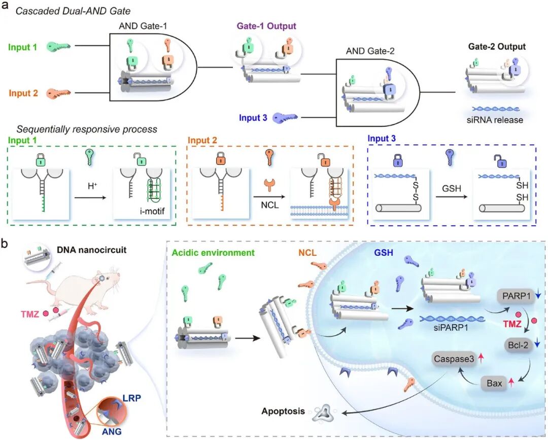
Figure 1.
Design and therapeutic application of the cascaded dual-AND logic DNA nanocircuit. (a) Schematic illustration of the DNA nanocircuit programmed with dual-AND logic gates. The first AND gate responds to extracellular acidic pH (Input 1) and membrane-overexpressed NCL (Input 2). The co-occurrence of these two tumor-specific signals triggers conformational rearrangement to generate a molecular output. This output (Gate-1 output) collaborates with intracellular GSH (Input 3) to activate the second AND gate. (b) In vivo application in the GBM mouse model. After crossing the blood–brain barrier via the binding of Angiopep-2 (ANG) to low-density lipoprotein receptor-related protein (LRP, which is highly expressed on both brain capillary endothelial cells and GBM cells), the nanocircuit sequentially responds to tumor-specific signals (acidic pH, NCL, and GSH). This enables precise siPARP1 release and gene silencing, thereby overcoming TMZ resistance and inducing prominent tumor cell apoptosis.
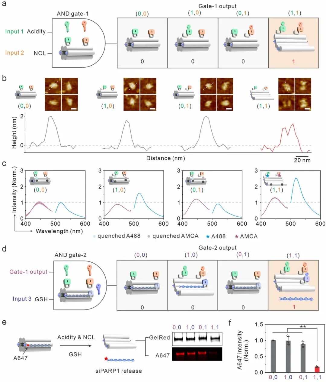
Figure 2.
In vitro validation of the activation of the cascaded dual-AND logic gates. (a) The first logic gate under four distinct input conditions: (0,0), (0,1), (1,0), and (1,1), corresponding to the absence or presence of acidic pH (Input 1) and NCL (Input 2), respectively. The output signal reads “1” only when both inputs are present. (b) AFM images showing the morphology of the nanocircuit under the four conditions (scale bar, 20 nm; full-field view in Figure S2). (c) Fluorescence readouts of the DNA nanocircuit. AMCA (λex = 345 nm, λem = 445 nm); A488 (λex = 480 nm, λem = 520 nm). Fluorescence intensities are normalized to the (0,0) input. Data are presented as mean ± SEM, depicted by solid lines and shaded regions (n = 3). (d) Schematic of the second logic gate, activated by the Gate-1 output and GSH (Input 3), which triggers siPARP1 release. (e) Schematic illustration of the release of A647-labelled siPARP1 from the DNA nanocircuit and PAGE analysis under four input conditions. (f) Quantification of the A647 fluorescence intensity of the nanocircuit bands. A647 intensities are normalized to the (0,0) input. All data are presented as mean ± SEM (n = 3), **p < 0.01.
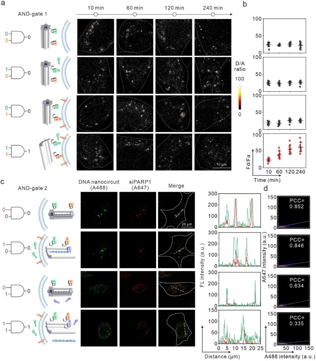
Figure 3.
Validation of the sequential logic behavior of the DNA nanocircuit in living cells. (a) Pseudocolored donor/acceptor images depicting the temporal response of the cell-surface AND gate-1 under different logic gate inputs. (b) Quantification of the D/A ratio from panel (a) based on n = 10 fluorescent spots at each time point, presented as mean ± SEM. (c) Colocalization of the DNA nanocircuit (A488-labeled, green) and siPARP1 (A647-labeled, red) for AND gate-2. The line profile shows the green and red fluorescence intensities along the dashed line in the merged image. (d) Colocalization plot of the cells in panel (c) and the corresponding Pearson correlation coefficient.
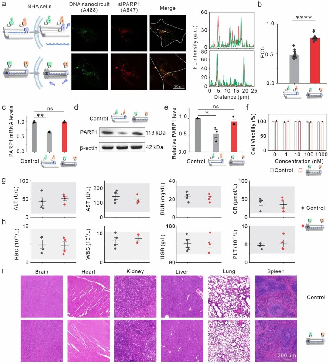
Figure 4.
Non-responsiveness and biosafety of the DNA nanocircuit in normal biological systems. (a) Colocalization of the DNA nanocircuit (A488-labeled, green) and siPARP1 (A647-labeled, red) in NHA cells incubated with AND gate-1-defective DNA nanocircuit (top) or cascaded dual-AND logic DNA nanocircuit (bottom). The line profile shows the green and red fluorescence intensities along the dashed line in the merged image. (b) Pearson correlation coefficients from 15 cells for each condition. (c) Relative PARP1 mRNA levels determined by qPCR under the two siPARP1 release conditions (n = 3). (d) Western blot analysis of PARP1 protein expression. (e) Densitometric quantification of panel (d) (n = 3). (f) Viability of NHA cells treated with DNA nanocircuit at different concentrations. (g, h) Serum biochemical indices (ALT, AST, BUN, CR) and hematological parameters (RBC, WBC, HGB, PLT) of mice after DNA nanocircuit injection. The gray shaded areas indicate standard reference ranges (n = 4). (i) Representative hematoxylin and eosin (H&E) staining of mouse brain, heart, kidney, liver, lung and spleen tissues after DNA nanocircuit administration. All data are presented as mean ± SEM. ns, not significant; *p < 0.05, **p < 0.01, ***p < 0.0001.
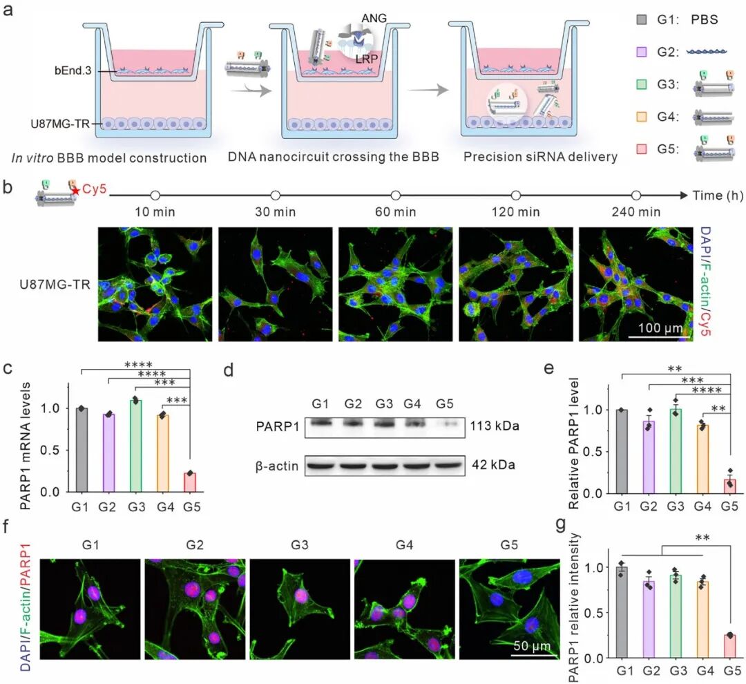
Figure 5.
In vitro evaluation of targeted PARP1 gene silencing by the DNA nanocircuit. (a) Schematic of the BBB Transwell system, with bEnd.3 cells cultured in the upper chamber and U87MG-TR cells in the lower chamber. The DNA nanocircuit crosses the upper barrier via ANG and is activated to release siPARP1 in the lower chamber. (b) Confocal imaging of cellular uptake of Cy5-labeled DNA nanocircuit by lower-chamber cells at indicated time points. (c) qPCR quantification of PARP1 mRNA expression under different treatments (n = 3). (d, e) Western blot detection of PARP1 protein (d) and corresponding densitometric analysis (e) (n = 3). (f) IF imaging of PARP1 expression in U87MG-TR cells and (g) quantitative analysis after treatment. All data are presented as mean ± SEM. **p < 0.01, ***p < 0.001, ****p < 0.0001.
Summary
siRNA therapy suffers from off‑target effects, and precise delivery is therefore essential. Herein, we engineered a DNA nanocircuit containing cascaded dual AND logic gates that sequentially respond to three tumor‑associated signals: extracellular acidic pH, overexpressed cell‑membrane nucleolin, and intracellular glutathione. The coexistence of acidic pH and nucleolin activates the first gate, triggering conformational rearrangement and generating a molecular output. This output, together with glutathione as the second input, activates the second gate to initiate siRNA release. In a temozolomide‑resistant glioblastoma model, the nanocircuit enables highly selective release of siPARP1, efficiently silences PARP1, reverses drug resistance and mitigates off‑target toxicity.
This work introduces multi‑signal logic computation into nucleic acid delivery, where drug release requires simultaneous satisfaction of multiple tumor hallmarks, thereby substantially improving selectivity at both spatial and molecular levels. The recognition modules can be replaced in future studies to accommodate different combinations of tumor biomarkers or alternative siRNA targets. The modular design of cascaded gates allows tuning of signal sequence and activation thresholds. This strategy is extendable to other nucleic acid agents requiring spatiotemporally controlled activation, such as mRNA and gene‑editing components. Further optimization is still needed regarding the in vivo stability and large‑scale synthesis cost of DNA circuits. Nevertheless, this logic‑gated framework provides a programmable and versatile platform for smart nucleic acid delivery.
Reference
DOI: 10.1002/advs.77332
We specialize in custom nano-medical functional materials. We conduct targeted development to tailor structure, particle size and surface modification for your experiments. Full characterization, iterative optimization and technical support are available for targeted delivery, bioimaging and nanomedicine studies. Send your evaluation plans to us.
Engineer Zhang: 17715390137
Engineer Chen: 18914047343