已传文件:photo/1793667293.png
 Metabolic reprogramming is one of the hallmark features of cancer, making the modulation of these pathways an attractive therapeutic strategy. However, clinical efficacy is frequently limited by compensatory metabolic networks and inefficient tumor targeting. Herein, we develop a catalytic nanogel to reprogram the immunosuppressive metabolic network for activatable cancer photoimmunotherapy. This nanogel (termed OMST) is fabricated by crosslinking oxidized hyaluronic acid with methylthioadenosine (MTA) catabolic enzyme (methylthioadenosine phosphorylase, MTAP), an MTA anabolic inhibitor (spermidine synthase inhibitor, SRMi), and a photosensitizer (TAPP) via acid-cleavable imine bonds. OMST can specifically dissociate and activate MTAP and SRMi only within the acidic tumor microenvironment (TME), enabling synergistic bidirectional metabolic reprogramming to promote MTA degradation and suppress MTA production, respectively. This bidirectional MTA depletion strategy remodels the immunosuppressive TME, inhibits pro-tumor M2 macrophages, facilitates antigen presentation by dendritic cells, and boosts the proliferation of cytotoxic T lymphocytes. Upon local irradiation, TAPP generates reactive oxygen species to trigger immunogenic cell death, which synergizes with MTA metabolic reprogramming to reinvigorate anti-tumor immunity. In vitro and in vivo experiments demonstrate that OMST efficiently suppresses tumor growth, metastasis and recurrence. Collectively, this study presents a precise and multidimensional metabolic reprogramming strategy for cancer therapy.

This study was published in Journal of Controlled Release under the title “Catalytical nanogels reprogram immunosuppressive metabolic networks for activatable cancer photo-immunotherapy”.
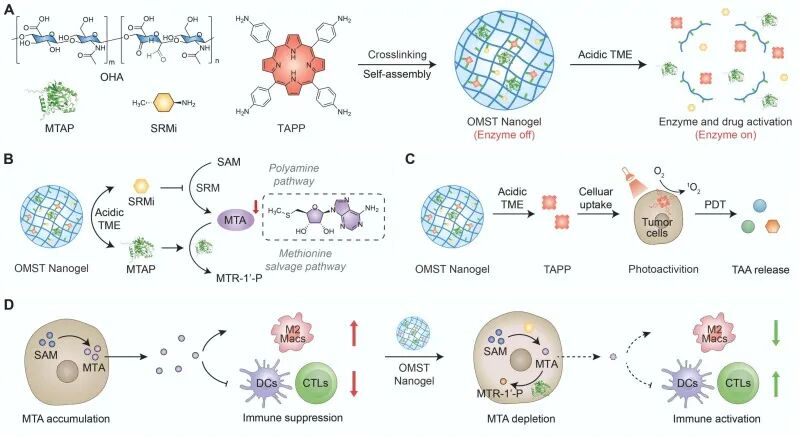
Figure 1
Schematic illustration of catalytic nanogels (OMST) that reprogram immunosuppressive metabolic networks for activatable cancer photoimmunotherapy. (A) Synthesis of OMST and its activation mediated by the acidic tumor microenvironment (TME). (B) Schematic showing the mechanism of bidirectional MTA metabolic reprogramming via MTAP-mediated catabolism and SRMi-inhibited anabolism. (C) OMST-mediated photodynamic therapy triggers ¹O₂ generation, immunogenic cell death (ICD), and the release of tumor-associated antigens. (D) OMST-mediated MTA depletion suppresses pro-tumor M2 macrophages, facilitates antigen presentation by DCs, and enhances the proliferation of CTLs to reprogram the immunosuppressive TME.
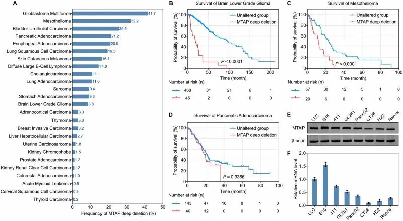
Figure 2
Statistics of MTAP deletion across pan-cancer cohorts. (A) Frequencies of deep MTAP deletions in human cancers. Data were obtained from cBioPortal for Cancer Genomics (http://www.cbioportal.org). Overall survival probabilities of (B) brain lower-grade glioma, (C) mesothelioma and (D) pancreatic adenocarcinoma. (E) Western blot and (F) RT-qPCR analyses of MTAP levels in several mouse tumor cell lines. Survival data were retrieved from The Cancer Genome Atlas, and survival analysis was performed using the log-rank (Mantel‑Cox) test.
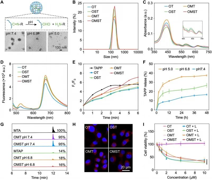 Figure 3
Synthesis and characterization of OMST for activatable cancer photoimmunotherapy. (A) Schematic illustration of acid-triggered nanogel dissociation in the acidic TME and TEM images of OMST in PBS solutions (pH 7.4, pH 6.8 and pH 5.0). (B) DLS profiles, (C) UV–Vis absorption spectra, and (D) fluorescence spectra of OT, OST, OMT and OMST in PBS. (E) ¹O₂ generation profiles of TAPP, OT, OST, OMT and OMST ([TAPP] = 20 μmol/L) under 670 nm laser irradiation (30 mW/cm²) in PBS. (F) Cumulative TAPP release profiles of OMST at pH 7.4, 6.8 or 5.0 over 48 h (n = 3). (G) HPLC chromatograms of MTA after OMT and OMST ([MTAP] = 1 μg/mL) were incubated with MTA (3 μmol/L) in PBS at pH 7.4 or 6.8 for 48 h. (H) Confocal fluorescence images of CT26 cells incubated with OT, OST, OMT and OMST ([TAPP] = 10 μmol/L) for 12 h. Blue and red fluorescence correspond to DAPI and TAPP, respectively. (I) Cell viability of CT26 cells after exposure to different concentrations of OT, OST, OMT and OMST for 24 h, with or without 670 nm laser irradiation (30 mW/cm², 6 min). Comparison of OMST at 10 μmol/L TAPP with versus without laser irradiation: ****p < 0.0001. Statistical significance in (I) was calculated using a two-tailed Student’s t-test.
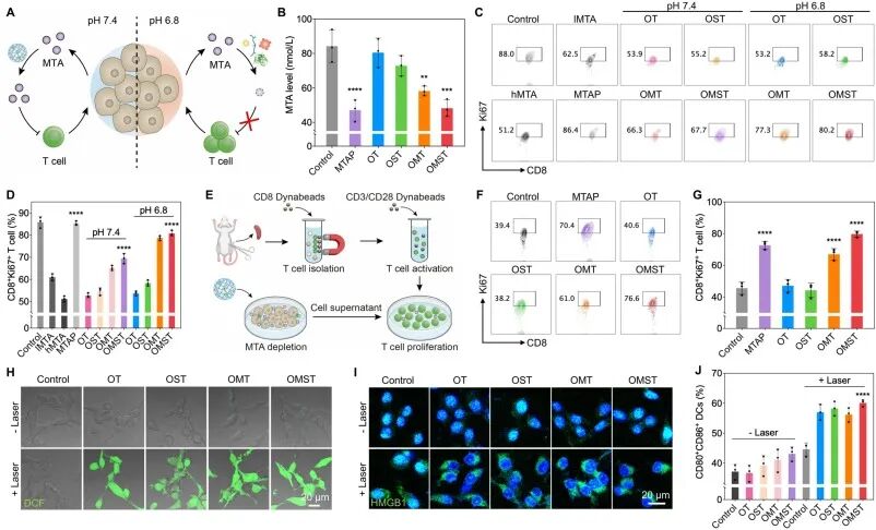
Figure 4
OMST-mediated activatable cancer photoimmunotherapy in vitro. (A) Schematic illustration of OMST-mediated MTA reprogramming and T cell proliferation in the acidic TME. (B) MTA levels determined by HPLC after CT26 cells were incubated with MTAP, OT, OST, OMT and OMST ([TAPP] = 20 μmol/L or [MTAP] = 0.1 μg/mL) for 12 h. MTAP vs control: p < 0.0001, OMT vs control: p < 0.01, OMST vs control: p < 0.001. (C) Flow cytometry and (D) quantitative analysis of proliferating CD8⁺Ki67⁺ T cells after 48 h incubation with MTA, MTAP, OT, OST, OMT and OMST (n = 3). lMTA: low MTA (125 μg/mL), hMTA: high MTA (250 μg/mL). OMST (pH 7.4 or pH 6.8) vs hMTA: ****p < 0.0001. (E) Schematic diagram showing the workflow of in vitro MTA depletion, T cell isolation, activation and proliferation assays. (F) Flow cytometry and (G) quantitative analysis of proliferating CD8⁺Ki67⁺ T cells after 48 h incubation with supernatants from differently treated CT26 cells (n = 3). MTAP, OMT or OMST vs control: ****p < 0.0001. (H) Confocal fluorescence images of CT26 cells incubated with OT, OST, OMT and OMST ([TAPP] = 20 μmol/L) for 12 h, stained with H2DCFDA, with or without 670 nm laser irradiation (30 mW/cm², 6 min). Green fluorescence indicates DCF. (I) Confocal fluorescence images of nuclear HMGB1 expression in CT26 cells incubated with OT, OST, OMT or OMST ([TAPP] = 20 μmol/L) for 12 h, with or without 670 nm laser irradiation (30 mW/cm², 6 min). (J) Quantification of mature DCs (CD80⁺CD86⁺) after BMDCs were co-cultured with supernatants from variously treated CT26 cells for 12 h (n = 3). OMST vs control: ****p < 0.0001. Statistical significance in (B), (D), (G) and (J) was calculated by one-way analysis of variance (one-way ANOVA) followed by Tukey’s post-hoc test. Data are presented as mean ± SD.
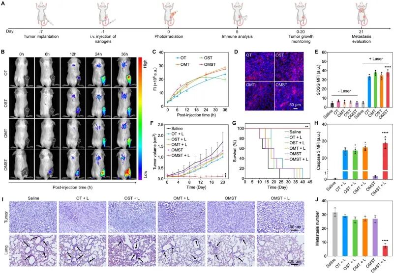
Figure 5
OMST-mediated activatable cancer photoimmunotherapy in vivo. (A) Schematic timeline showing the establishment of the CT26 tumor model and the regimen of subsequent OMST-mediated activatable cancer photoimmunotherapy. (B) In vivo fluorescence images of tumors in CT26 tumor-bearing mice at 0, 6, 12, 24 or 36 h after intravenous injection of OT, OST, OMT or OMST (injection volume: 200 μL, [TAPP] = 1 mmol/L), and (C) quantitative fluorescence intensity (n = 3). (D) Confocal fluorescence images of tumor tissues harvested from CT26 tumor-bearing mice at 36 h post-injection. Blue fluorescence indicates DAPI, and red fluorescence indicates TAPP. (E) Mean fluorescence intensity of SOSG in tumor tissues of CT26 tumor-bearing mice receiving different treatments, with or without 670 nm laser irradiation (30 mW/cm², 6 min) (n = 5). OMST+L vs saline: ****p < 0.0001. (F) Tumor growth curves recorded over the 21-day monitoring period following different treatments (n = 5). OMST+L vs saline on day 20: ***p < 0.001. (G) Kaplan–Meier survival curves of tumor-bearing mice subjected to various treatments (n = 5). OMST+L vs saline: **p < 0.01. (H) Quantification of caspase-3 expression (n = 5). OMST+L vs saline: ****p < 0.0001. (I) Histological H&E staining of tumor tissues collected on day 14 and lung tissues collected on day 21. (J) Count of metastatic nodules in histological images of lung tissues from CT26 tumor-bearing mice after different treatments (n = 3). OMST+L vs saline: ****p < 0.0001. Statistical significance in (E), (H) and (J) was determined by one-way analysis of variance (one-way ANOVA) followed by Tukey’s post-hoc test. Statistical significance in (F) was determined by a two-tailed Student’s t-test. Statistical significance in (G) was determined by the log-rank test. Data are presented as mean ± SD.
Summary
Compensatory feedback within tumor metabolic networks and inadequate tumor targeting restrict the therapeutic outcomes of metabolic interventions. Herein, we engineered acid-responsive catalytic nanogels crosslinked via imine bonds from oxidized hyaluronic acid, methylthioadenosine-degrading enzyme, an inhibitor of methylthioadenosine biosynthesis, and a photosensitizer. The nanogels dissociate and release the two metabolic regulatory components exclusively in the acidic tumor microenvironment, enabling bidirectional depletion of methylthioadenosine by accelerating its degradation and blocking its production. Such metabolic reprogramming suppresses M2-like macrophages, facilitates antigen presentation by dendritic cells, and boosts the proliferation of cytotoxic T lymphocytes. Upon local irradiation, the photosensitizer generates reactive oxygen species to induce immunogenic cell death, which synergizes with metabolic modulation to activate anti-tumor immunity and suppress tumor growth, metastasis and recurrence in vivo.
This work integrates the “bidirectional depletion” metabolic intervention and photoimmunotherapy within a single acid-responsive nanogel, circumventing compensatory pathways triggered by monotargeted metabolic inhibition. In the future, other tumor-specific metabolic nodes can be screened to develop analogous bidirectional regulatory combinations. The acid-cleavable imine bond design can be generalized to other delivery systems requiring tumor microenvironment-triggered activation. This strategy is also applicable to other tumor types with aberrant methylthioadenosine metabolism. The spatiotemporal controllability of local irradiation makes it suitable for superficial lesions or lesions accessible via endoscopy. Although the long-term impacts of sustained metabolic reprogramming on normal cells remain to be further evaluated, this multidimensional design provides a practical nanoplatform for synergistic metabolic–immunotherapy.
Reference
DOI: 10.1016/j.jconrel.2026.115384
We specialize in custom nano-medical functional materials. We conduct targeted development to tailor structure, particle size and surface modification for your experiments. Full characterization, iterative optimization and technical support are available for targeted delivery, bioimaging and nanomedicine studies. Send your evaluation plans to us.
Engineer Zhang: 17715390137
Engineer Chen: 18914047343