已传文件:photo/1793671560.png
 Multi-omics analysis indicates that enhanced oxidative phosphorylation (OXPHOS) is associated with breast cancer bone metastasis (BCBM). To target this metabolic vulnerability, we engineered SPMMPA, a bone-targeted nanozyme composed of a Prussian blue core, a disulfide-bridged mesoporous silica interlayer, and a manganese oxide shell, functionalized with polyvinylamine, alendronate, and the Ser-Asp-Ser-Ser-Asp peptide. Its acid-boosted catalytic activity promotes reactive oxygen species generation and glutathione depletion, accompanied by mitochondrial dysfunction, reduced oxaloacetate and adenosine triphosphate production, and downregulation of peroxisome proliferator-activated receptor γ coactivator 1α. The release of alendronate and silicon helps attenuate osteoclast activity and preserve bone architecture. Near-infrared irradiation further augments therapeutic efficacy via mild photothermal heating below 48 °C. In the BCBM mouse model, SPMMPA suppresses tumor growth and alleviates osteolysis with no detectable obvious toxicity throughout the observation period. These findings support a pH-responsive catalytic strategy that couples disruption of tumor energy metabolism with the preservation of bone homeostasis.

This study was published in Advanced Materials under the title “A Novel pH-Activatable Nanozyme for Treating Breast Cancer Bone Metastasis by Regulating Cancer Cell Metabolic Reprogramming and the Tumor Microenvironment”.
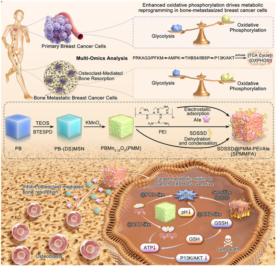
Figure 1
Schematic illustration of the design of SPMMPA nanozyme and the relationship among its catalytic activity, tumor energy metabolism and bone homeostasis for breast cancer bone metastasis.
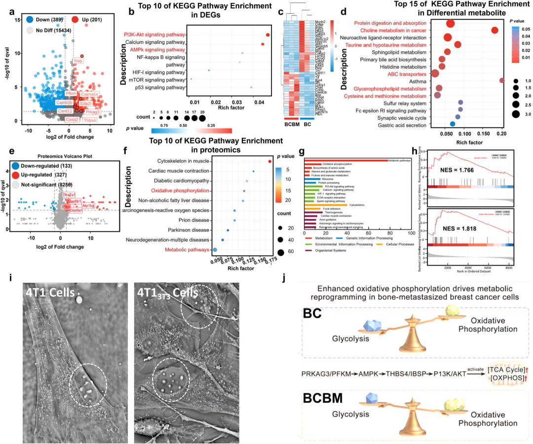
Figure 2
Metabolic reprogramming enhances OXPHOS in breast cancer bone metastasis. Volcano plot of DEGs (red: upregulated genes; blue: downregulated genes; gray: non-significant DEGs) (a). Energy-metabolism-related DEGs from KEGG pathway enrichment analysis (top-ranked pathways by p value) (b). Heatmap of energy-metabolism-related DEGs (red: high expression; blue: low expression) (c). KEGG pathway enrichment analysis for different metabolites (top-ranked pathways by p value) (d). Volcano plot of proteomic data (red: upregulated proteins; blue: downregulated proteins; gray: non-significant proteins) (e). Proteomic KEGG pathway enrichment analysis involving energy metabolism (top-ranked pathways by p value) (f). Differential distribution of proteins across different bioactivities (g). Gene set enrichment analysis (GSEA) (h). Label-free live-cell images; dashed lines indicate regions of interest (i). Proposed mechanism of oxidative phosphorylation system reprogramming in 4T1 cells during breast cancer bone metastasis (j).
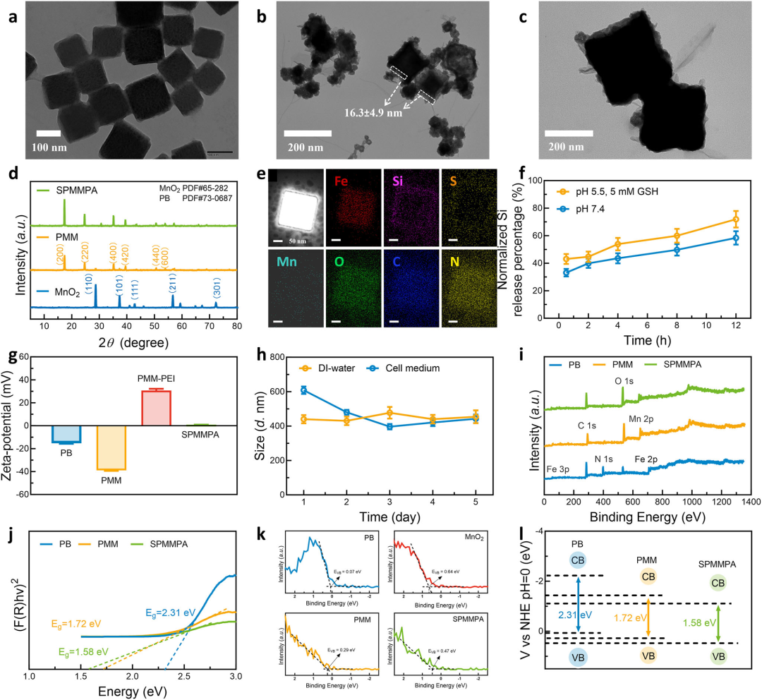
Figure 3
Morphology, microstructure and physicochemical properties of NPMM and SPMMPA nanozymes. TEM images of PMM (a) and SPMMPA (b and c). XRD patterns of MnO₂, PMM and SPMMPA (d). HAADF‑STEM image and elemental mapping images of SPMMPA (e). ICP‑MS quantification of released Si from SPMMPA in PBS at pH 5.5 (with 5 mM GSH) versus pH 7.4 (without GSH) (f). Zeta potentials of PB, PMM, PMM‑PEI and SPMMPA (g). Size stability of SPMMPA in deionized water and RPMI 1640 medium (pH 7.4, 10 mM, 10% FBS, 37 °C) (h). XPS survey spectra (i), plots of transformed Kubelka–Munk function versus photon energy (j), and band structures of PB, PMM and SPMMPA (l). High‑resolution XPS spectra of PB, MnO₂, PMM and SPMMPA (k).
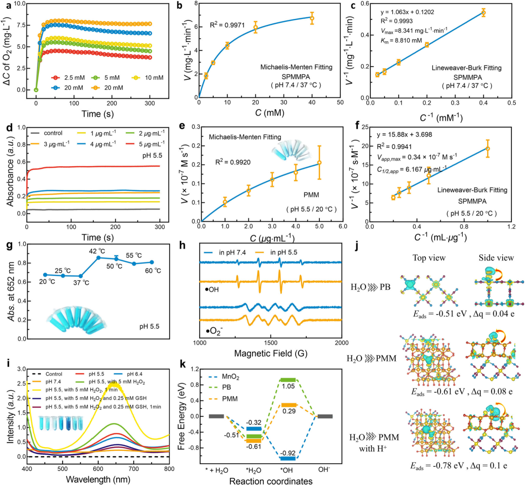
Figure 4
Enzymatic activity investigation of SPMMPA. Variation in H₂O₂ concentration in aqueous suspensions of SPMMPA with altered SPMMPA concentrations (PMM concentration fixed at 25 μg·mL⁻¹) in the presence of H₂O₂ at approximately 37 °C and pH 7.4 (a). Michaelis–Menten kinetic analysis (b) and Lineweaver–Burk plot (c) for the CAT-like activity of SPMMPA using H₂O₂ as the substrate. Time-dependent absorbance changes at 652 nm upon TMB oxidation in PBS buffer (pH 5.5) with different concentrations of SPMMPA at 20 °C (d). Empirical catalyst–concentration–reaction parameters for the OXD-like activity of SPMMPA with H₂O₂ and O₂ as substrates (e and f). Absorbance changes at 652 nm during TMB oxidation catalyzed by PMM (4 μg·mL⁻¹) in PBS (pH 5.5) at different temperatures (g). EPR spectra showing the generation of •OH and •O₂⁻ by PMM at different pH values (h). UV–Vis spectra of TMB oxidation catalyzed by PMM (4 μg·mL⁻¹) in aqueous dispersion with H₂O at 20 °C in PBS buffer (pH 5.5, 6.4 or 7.4), with or without 5 mM H₂O₂ and/or 0.25 mM GSH after 0 min or 1 min of reaction (i). Optimized adsorption charge difference distribution of H₂ molecules on PB, MnO₂ and PMM with H₂ loading (j). Free energy diagrams for PB, MnO₂ and the PMM model during the OXD-like catalytic process (k).
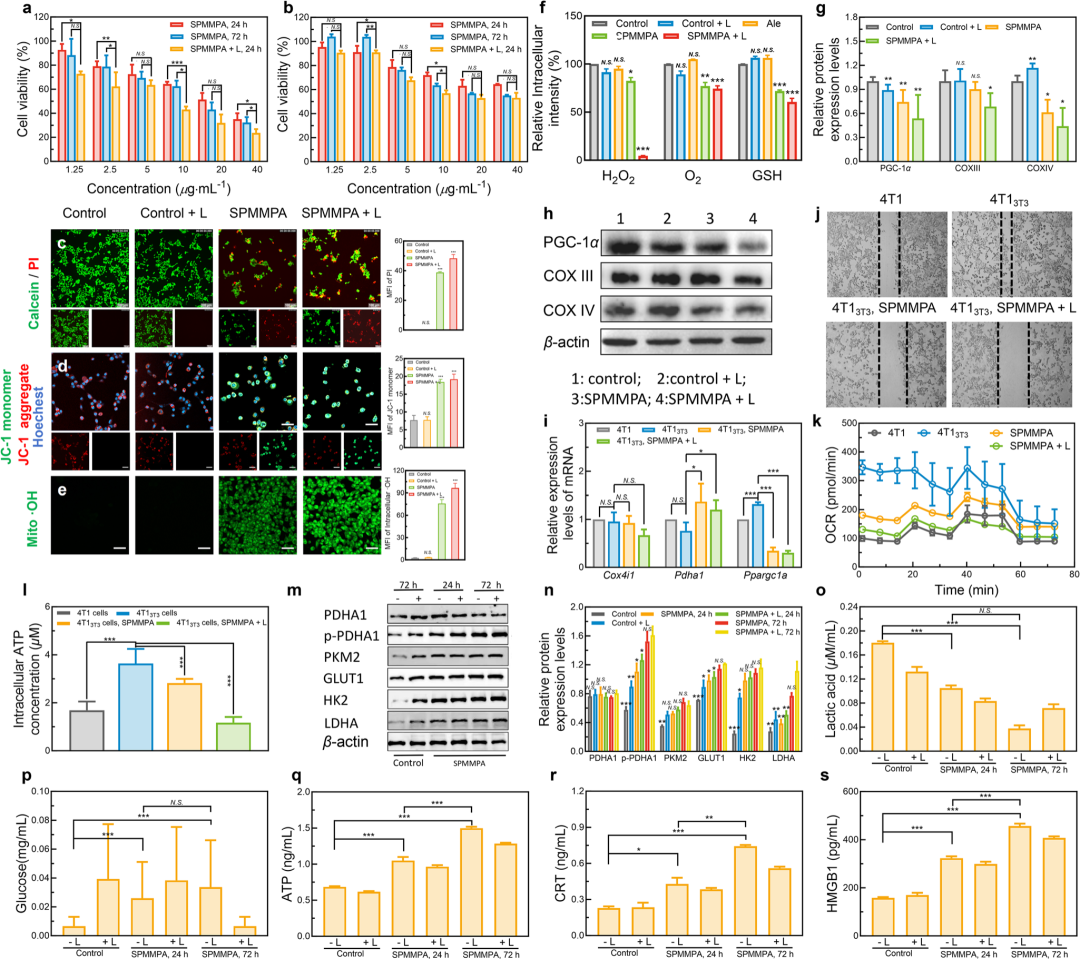
Figure 5
SPMMPA suppresses the malignant behaviors of tumor cells in vitro. Cell viability of 4T1 cells (a) and 3T3 cells (b) incubated with SPMMPA for 24 h or 72 h with or without 808 nm laser irradiation (1.25 W cm⁻²) for 5 min under normoxic conditions. CLSM images of Calcein‑AM/PI double‑stained 4T1 cells after various treatments (c). Scale bar: 100 μm. CLSM images of JC‑1‑stained 4T1 cells after different treatments (d). Red JC‑1 aggregates indicate mitochondria with normal membrane potential, and green JC‑1 monomers indicate mitochondria with depolarized membranes (L: 808 nm laser irradiation (1.25 W cm⁻²) for 5 min). Scale bar: 50 μm. CLSM images of •OH in 4T1 cells after different treatments (e). Scale bar: 50 μm. Relative intracellular H₂O₂ and GSH concentrations in 4T1 cells after different treatments (SPMMPA concentration: 5 μg·mL⁻¹), without or with 808 nm laser irradiation (1.25 W cm⁻², 5 min), measured by a microplate reader (f). Western blot analysis of PGC‑1α, COX III and COX IV expression in 4T1 cells incubated with SPMMPA with or without laser irradiation (h). The relative expression levels of proteins were normalized to β‑actin as the internal reference.
mmary
Multi‑omics analysis reveals enhanced oxidative phosphorylation in breast cancer bone metastasis. Targeting this metabolic vulnerability, we designed a bone‑targeted nanozyme termed SPMMPA. This nanozyme consists of a Prussian blue core, a disulfide‑bridged mesoporous silica interlayer and a manganese oxide shell, and is modified with PEI, alendronate and SDSSD peptide to enhance targeted accumulation within bone tissue. The acidic microenvironment boosts its catalytic activity, facilitating reactive oxygen species production and glutathione depletion. This process is accompanied by impaired mitochondrial function, decreased oxidative phosphorylation and ATP levels, as well as downregulation of PGC‑1α. Meanwhile, the release of alendronate and silicon helps reduce osteoclast activity and preserve bone architecture, and near‑infrared photothermal treatment further amplifies the therapeutic effects.
In the mouse model of bone metastasis, SPMMPA suppresses tumor progression and mitigates osteolysis with no obvious toxicity observed during the monitoring period. These results demonstrate that the pH‑responsive catalytic strategy integrates perturbation of tumor energy metabolism and protection of bone homeostasis within a single platform. Future work should focus on dissecting the respective contributions of SDSSD peptide and alendronate to bone targeting, clarifying the causal links among catalytic activity, metabolic suppression and immune alterations. Additional validation of long‑term biosafety and evaluation in diverse bone metastasis models are also required to define the applicable scope of this bone‑targeted catalytic strategy.
Reference
DOI: 10.1002/adma.75432
We specialize in custom nano-medical functional materials. We conduct targeted development to tailor structure, particle size and surface modification for your experiments. Full characterization, iterative optimization and technical support are available for targeted delivery, bioimaging and nanomedicine studies. Send your evaluation plans to us.
Engineer Zhang: 17715390137
Engineer Chen: 18914047343