已传文件:photo/1794376932.png
 Abstract
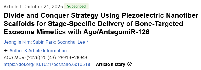
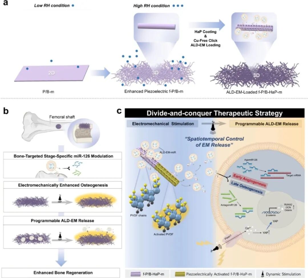
Bone regeneration is regulated by spatiotemporally ordered angiogenic and osteogenic signals. Most existing scaffolds rely on passive release and fail to match the stages of healing. Herein, we fabricate a multifunctional piezoelectric nanofibrous scaffold for mechanically triggered, temporally sequenced delivery of bone‑targeted exosome mimetics loaded with either AgomiR‑126 or AntagomiR‑126. The three‑dimensional piezoelectric structure generates electromechanical activation under physiological loading, coupling mechanical stimulation with therapeutic release. ALD‑functionalized EM enables bone‑selective localization. AgomiR‑126 preferentially promotes early angiogenesis and progenitor cell recruitment, whereas AntagomiR‑126 facilitates later osteogenesis. In vivo results demonstrate that this system improves bone repair, primarily by establishing a mechanically gated, temporally programmed miRNA delivery framework.
01
Research Background
Bone healing requires sequential coordination of angiogenesis, inflammation resolution and osteogenesis. Single passive scaffolds struggle to adapt to these dynamic stages. Piezoelectric materials convert mechanical stimuli into electrical signals, yet most systems only focus on signal output or osteoconduction, lacking dynamic regulation of therapeutic activity. miRNAs can modulate multi‑gene networks, but their therapeutic application is limited by insufficient temporal control. miR‑126 promotes angiogenesis while inhibiting osteogenesis; AgomiR and AntagomiR exert opposite functions. EV‑based delivery offers distinct advantages, yet it suffers from poor targeting, uncontrolled release and off‑target accumulation. Integrating mechanically responsive release with stage‑specific miRNA functions forms the starting point of this study.
02 Main Content
A high‑β‑phase PVDF matrix incorporated with BTO and MWCNT is electrospun under high‑humidity conditions to form a fluffy three‑dimensional piezoelectric scaffold. Hydroxyapatite coating is generated via mineralization in simulated body fluid, and mineralization is promoted through charge mediation to provide binding sites for ALD‑EM. EM is produced by extrusion of MSCs. Metabolic glycoengineering is used to introduce azide groups, followed by bioorthogonal conjugation of DBCO‑ALD to achieve bone targeting, and miRNAs are loaded by electroporation. Serving as a mechanically gated interface, the scaffold regulates EM release upon mechanical deformation, thereby coordinating early vascularization and late osteogenesis within a single system.
03 Study Design
Fiber morphology, crystal phase, piezoelectric output, mineralization quality, EM immobilization and mechanically responsive release were characterized. In vitro assays were performed to evaluate hMSC infiltration, proliferation, osteogenic and angiogenic markers, EM uptake, as well as changes in RUNX2 after YAP knockdown. A rat femoral cortical defect model was adopted in vivo. Scaffolds loaded with AgomiR‑126 or AntagomiR‑126 were compared, with mechanical stimulation applied at early or late time windows. Bone and vascular responses were assessed using micro‑CT, histology, immunohistochemistry, CD31/EMCN double staining and angiography.
04 Results
The fluffy scaffolds exhibit superior piezoelectric output, mineralization and cell infiltration. ALD‑EM is stably immobilized via bisphosphonate‑hydroxyapatite interactions. Mechanical stimulation facilitates the release of EM and miRNAs in a frequency‑dependent manner. In vitro, AgomiR‑126 upregulates angiogenic markers and suppresses osteogenic markers, whereas AntagomiR‑126 promotes osteogenesis. Mechanical stimulation enhances osteogenesis via the YAP/RUNX2 axis. In vivo, early stimulation favors angiogenesis induced by AgomiR‑126, while late stimulation facilitates AntagomiR‑126‑mediated osteogenesis and defect bridging with mild inflammatory responses.
05 Outlook
This study transforms scaffolds from passive carriers into mechanically gated therapeutic interfaces, highlighting the matching between release timing and biological stages. Future work should evaluate the long‑term retention and tissue responses of non‑degradable PVDF/BTO/MWCNT systems and validate the performance in more complex defects and large animal models. Other mechanosensitive pathways and bioelectrical mechanisms beyond YAP need to be dissected. Stimulation frequency, time windows and loading sequences can be further optimized. Degradable or hybrid piezoelectric systems, EM heterogeneity, loading control and selective intraskeletal cell delivery are also worthy of exploration.
Reference:
ACS
Nano
Pub Date: 2026‑11‑03
DOI: 10.1021/acsnano.6c10518
Jeong In Kim, Subin Park, Soonchul Lee
We specialise in custom development of functional nanomedicine materials. Materials can be tailored according to experimental requirements, with tunable structure, particle size and surface modification. Full characterization services are available. Solutions can be iteratively optimised with continuous technical support throughout the project. Our services support research projects including targeted delivery, bioimaging and nanotheranostics. Please feel free to share your specifications for solution evaluation.
Mr. Zhang: +86 17715390137
Mr. Chen: +86 18914047343