已传文件:photo/1794375524.png
 Mitochondrial transplantation represents an important therapeutic strategy to restore energy supply in patients with ischemic heart disease (IHD). However, its application is limited by the invasiveness of transplantation approaches and the loss of mitochondrial activity. Herein, we report a successful oral mitochondrial transplantation strategy for IHD treatment. Nitric oxide-releasing nanomotors are engineered on the mitochondrial surface to construct nanomotor-equipped mitochondria with chemotactic targeting toward injured cardiac tissue via nanomotion. These nanomotorized mitochondria are encapsulated within enteric capsules to protect them from gastric acid erosion. After oral administration, the mitochondria are released in the intestine, rapidly taken up by intestinal cells and secreted into the bloodstream, enabling delivery to the injured heart tissue. The modulation of the disease microenvironment by nanomotorized mitochondria not only facilitates rapid uptake and high retention of transplanted mitochondria in injured cardiomyocytes, but also preserves the high activity of grafted mitochondria. Furthermore, in vivo results from IHD animal models demonstrate that accumulated nanomotorized mitochondria in damaged cardiac tissue can regulate cardiac metabolism at the transcriptional level, thereby halting IHD progression. This strategy holds the potential to transform therapeutic regimens for IHD.

This study was published in Nature Nanotechnology under the title “Oral mitochondrial transplantation using nanomotors to treat ischaemic heart disease”.
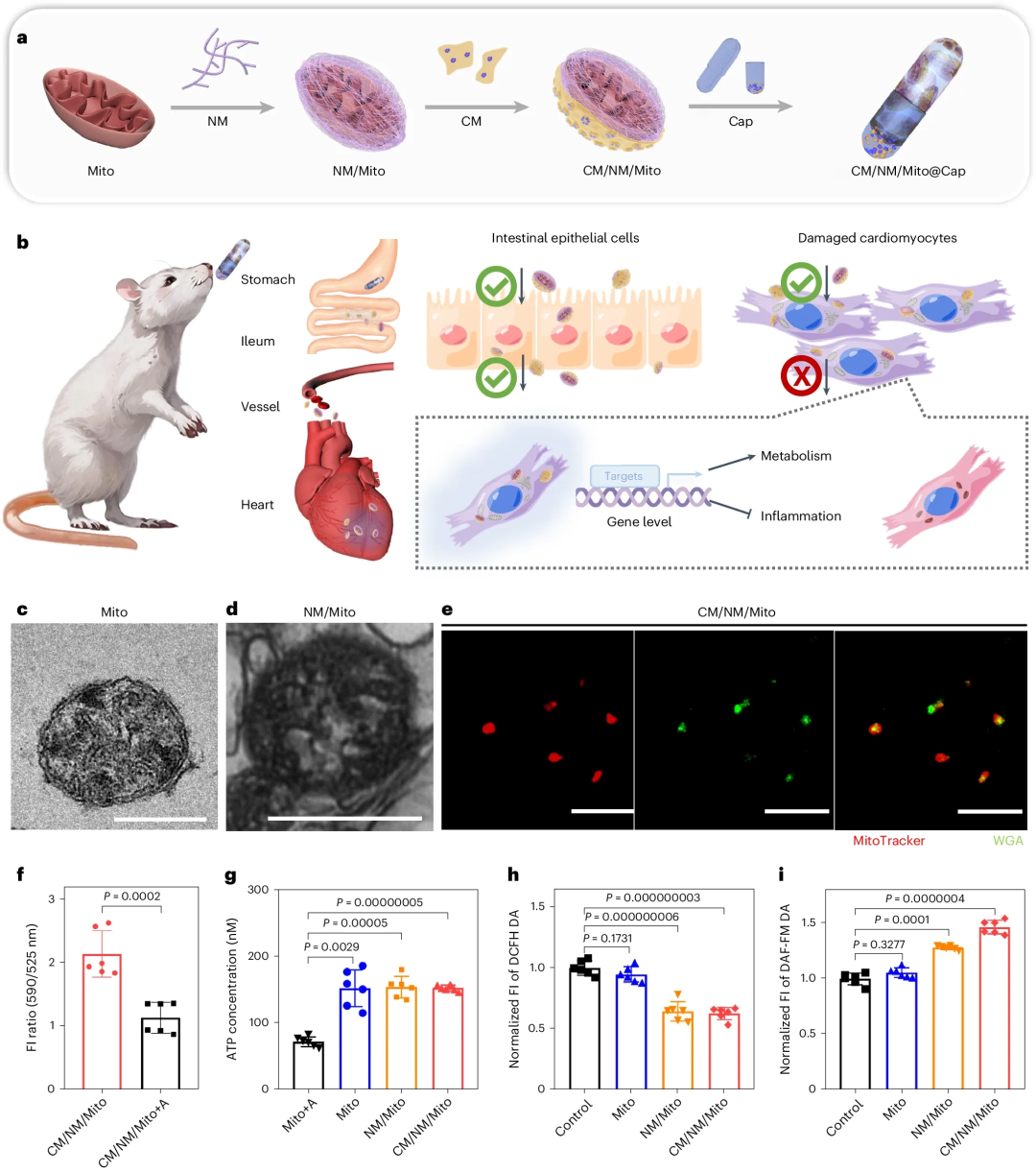
Figure 1
a, Fabrication of nanomotor and cell membrane modification, as well as compartmental design of the capsule.
b, In vivo delivery and therapeutic pathway of CM/NM/Mito@Cap.
c, d, TEM images of unmodified mitochondria (c) and NM/Mito (d). Scale bars, 500 nm.
e, CLSM images of CM/NM/Mito. MitoTracker, NM/Mito; WGA, CM. Scale bar, 10 μm.
f, Quantitative analysis of CM/NM/Mito after JC‑1 staining, with antimycin A‑treated CM/NM/Mito as control. n = 6 biologically independent samples. FI, fluorescence intensity.
g, ATP synthesis capacity of unmodified mitochondria, NM/Mito and CM/NM/Mito in the presence of substrates malate, glutamate and ADP. n = 6 biologically independent samples.
h, i, Flow cytometry quantification of ROS (h) and NO (i) levels in injured H9c2 cells after 24 h of incubation with different samples. n = 6 biologically independent cell samples; gating strategy is shown in Extended Data Fig. 2h.
Data are presented as mean ± s.d. Statistical significance was calculated using a two‑tailed unpaired Student’s t‑test in f, one‑way ANOVA followed by two‑tailed least‑significant‑difference (LSD) multiple comparison test in h, and two‑tailed Dunnett’s T3 multiple comparison test in g and i.
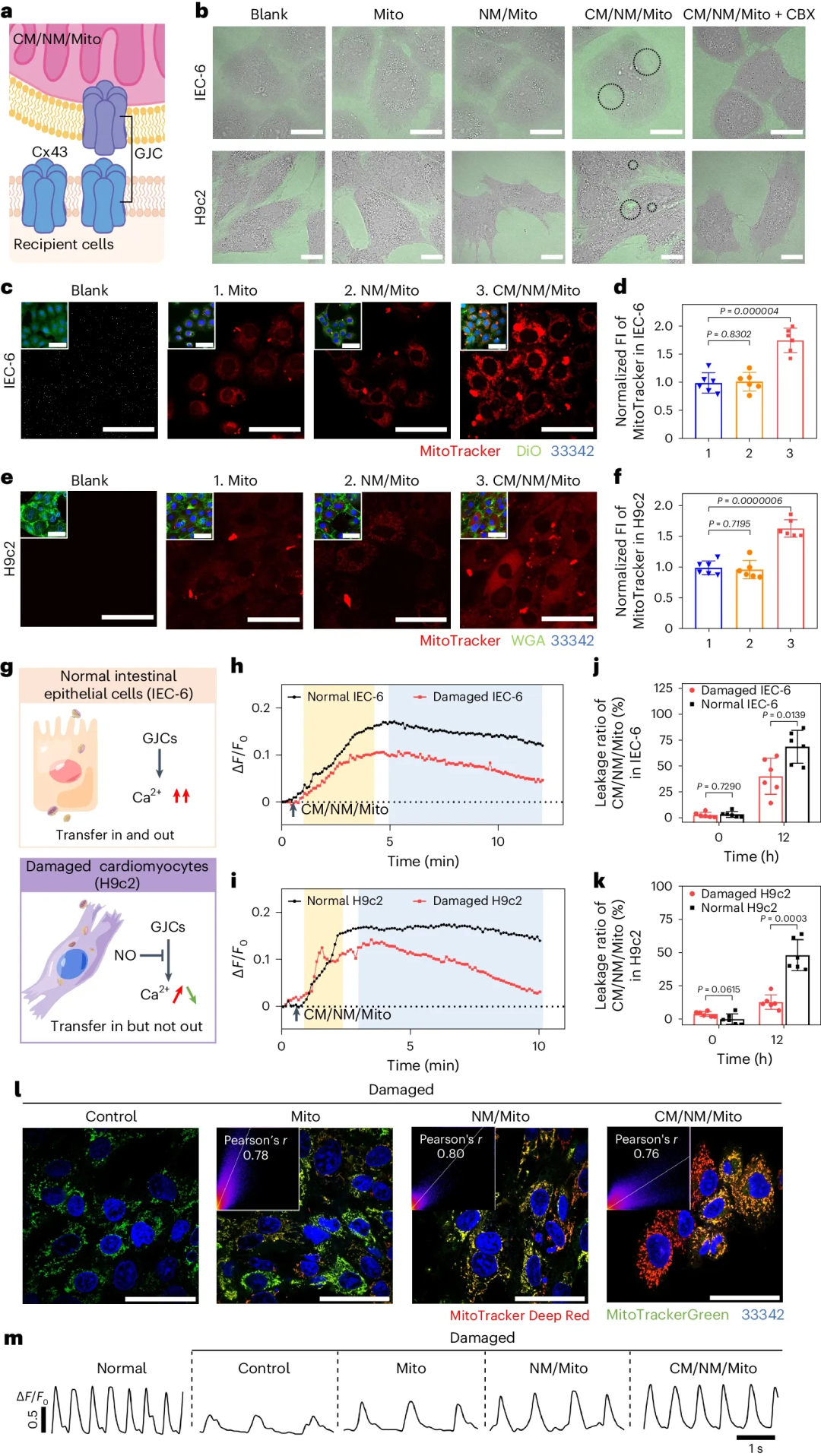
Figure 2
a, Schematic illustration of gap junction channel formation between CM/NM/Mito and recipient cells.
b, CLSM images showing gap junction channels formed between CM/NM/Mito and normal IEC‑6 and H9c2 cells in calcein‑containing medium. Black circles, calcein‑stained regions. Scale bar, 20 μm.
c–f, CLSM images and corresponding quantification of different samples in normal IEC‑6 (c, d) and H9c2 cells (e, f). MitoTracker, samples; DiO or WGA, cell membrane; Hoechst 33342, nucleus. Scale bars, 50 μm. (1) Unmodified mitochondria; (2) NM/Mito; (3) CM/NM/Mito. n = 6 biologically independent cell samples.
g, Mechanism for selective exocytosis of CM/NM/Mito by cells in different states.
h, i, Ca²⁺ level profiles in normal and injured IEC‑6 (h) and H9c2 cells (i).
j, k, Quantification of CM/NM/Mito leakage ratio in normal and injured IEC‑6 (j) and H9c2 cells (k). n = 6 biologically independent cell samples.
l, CLSM images and Pearson correlation coefficients for colocalization of endogenous mitochondria with different samples in injured H9c2 cells. MitoTracker Deep Red, samples; MitoTracker Green, endogenous mitochondria; Hoechst 33342, nucleus. Scale bar, 50 μm.
m, Calcium transients of primary cardiomyocytes after incubation with different samples for 24 h.
Data are presented as mean ± s.d. Statistical significance was calculated using one‑way ANOVA followed by two‑tailed LSD multiple comparison test in d and f, and two‑tailed unpaired Student’s t‑test in j and k.
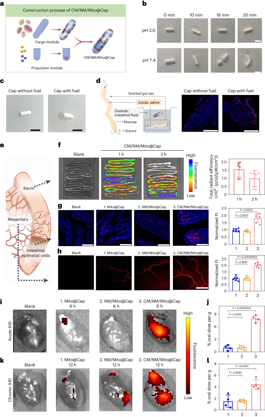
Figure 3
a, Schematic illustration of the assembly process of CM/NM/Mito@Cap.
b, Disintegration process of fuel‑loaded capsules in PBS. Scale bar, 5 mm.
c, Gas production of capsules with or without fuel in rat intestinal fluid. Scale bar, 5 mm.
d, Schematic of the everted intestinal sac and CLSM images of ileum sections. MitoTracker, CM/NM/Mito; DAPI, nucleus. Scale bar, 500 μm.
e, Schematic of observation sites.
f, Ex vivo imaging and quantification of intestinal tissues from rats after oral administration of CM/NM/Mito@Cap. n = 6 biologically independent animal samples.
g, h, CLSM images and quantification of ileum sections (g), and in vivo fluorescence images and quantification of mesenteric microvessels (h) from rats orally administered with (1) Mito@Cap, (2) NM/Mito@Cap and (3) CM/NM/Mito@Cap. MitoTracker, samples; DAPI, nucleus. Scale bar, 100 μm. n = 6 biologically independent animal samples.
i–l, Ex vivo cardiac imaging and cardiac delivery efficiency after oral administration of (1) Mito@Cap, (2) NM/Mito@Cap and (3) CM/NM/Mito@Cap in acute IHD models (i, j) and chronic IHD models (k, l). n = 4 biologically independent animal samples.
Data are presented as mean ± s.d. Statistical significance was calculated using one‑way ANOVA followed by two‑tailed Dunnett’s T3 multiple comparison test in g, and two‑tailed LSD multiple comparison test in h, j and l.
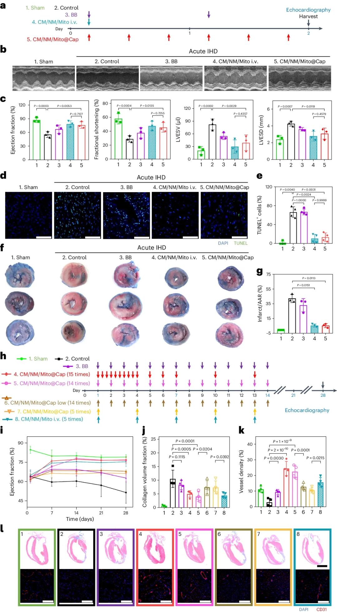
Figure 4
a, Schematic illustration of the assembly process of CM/NM/Mito@Cap.
b, Disintegration process of fuel‑loaded capsules in PBS. Scale bar, 5 mm.
c, Gas production of capsules with or without fuel in rat intestinal fluid. Scale bar, 5 mm.
d, Schematic of the everted intestinal sac and CLSM images of ileum sections. MitoTracker, CM/NM/Mito; DAPI, nucleus. Scale bar, 500 μm.
e, Schematic of observation sites.
f, Ex vivo imaging and quantification of intestinal tissues from rats after oral administration of CM/NM/Mito@Cap. n = 6 biologically independent animal samples.
g, h, CLSM images and quantification of ileum sections (g), and in vivo fluorescence images and quantification of mesenteric microvessels (h) from rats orally administered with (1) Mito@Cap, (2) NM/Mito@Cap and (3) CM/NM/Mito@Cap. MitoTracker, samples; DAPI, nucleus. Scale bar, 100 μm. n = 6 biologically independent animal samples.
i–l, Ex vivo cardiac imaging and cardiac delivery efficiency after oral administration of (1) Mito@Cap, (2) NM/Mito@Cap and (3) CM/NM/Mito@Cap in acute IHD models (i, j) and chronic IHD models (k, l). n = 4 biologically independent animal samples.
Data are presented as mean ± s.d. Statistical significance was calculated using one‑way ANOVA followed by two‑tailed Dunnett’s T3 multiple comparison test in g, and two‑tailed LSD multiple comparison test in h, j and l.
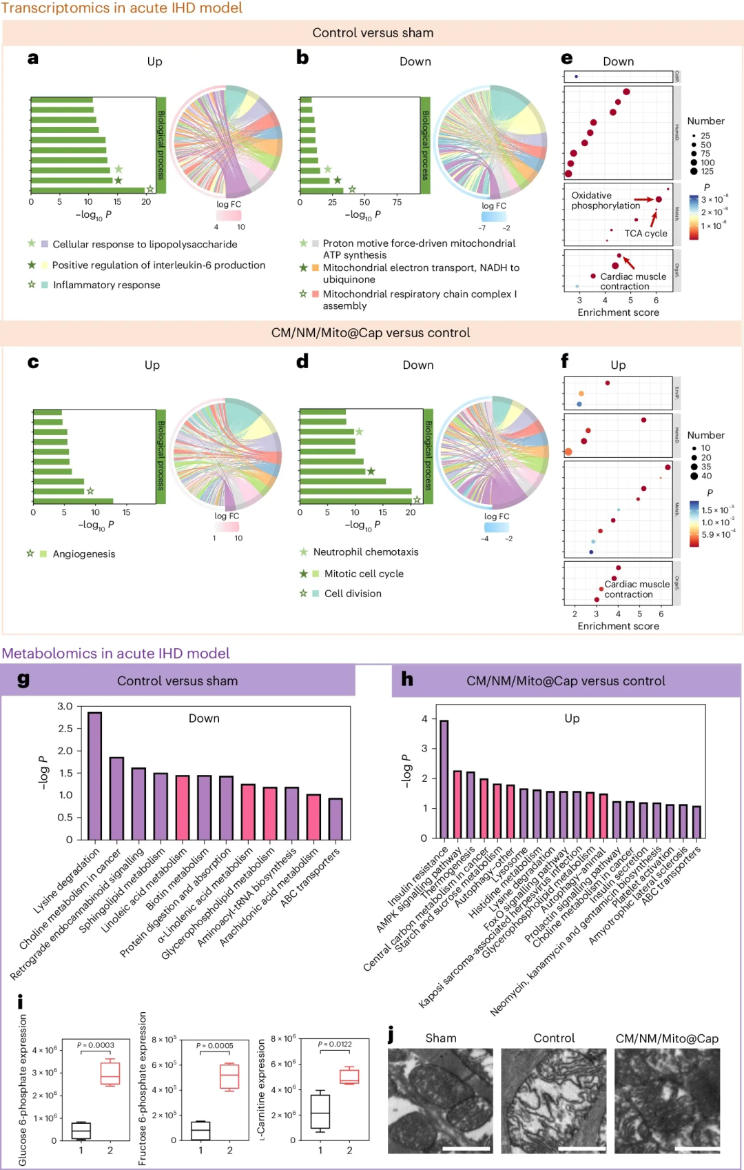
Figure 5
a–f, Transcriptomic analysis in the acute IHD model. GO enrichment analysis showing DEGs when comparing the control group versus the sham group (a,b), and the CM/NM/Mito@Cap group versus the control group (c,d), together with the corresponding KEGG pathway enrichment analysis (e,f). Histograms: the top ten significantly enriched biological processes. The horizontal axis denotes −log₁₀ P of pathways, and the vertical axis denotes GO terms. Chord diagrams: the top ten significantly enriched GO terms. The right semicircle shows the names of the ten GO terms, and the left semicircle shows the DEGs within these GO terms. The colour map represents gene fold changes (log₂ scale), and coloured ribbons connect genes to specific GO terms. Bubble plots: the top 20 significantly enriched KEGG terms. The horizontal axis denotes enrichment score, the vertical axis denotes descriptions, and bubble size denotes the number of enriched genes. CellP., Cellular Processes; HumaD., Human Diseases; Metab., Metabolism; OrgaS., Organismal Systems; EnvIP., Environmental Information Processing.
g–i, Untargeted metabolomic analysis in the acute IHD model: histograms of metabolic pathway enrichment analysis (g,h), and box plots of representative differential metabolites (including glucose 6‑phosphate, fructose 6‑phosphate and L‑carnitine), showing the median (line inside the box), interquartile range (box), 1.5 × interquartile range (whiskers), minimum and maximum values (i) ((1) Control; (2) Oral CM/NM/Mito@Cap).
j, TEM images of mitochondria in the heart. Scale bar, 1 μm.
In a–i, n = 3 biologically independent animal samples for the sham group, and n = 4 biologically independent animal samples for the control group and the oral CM/NM/Mito@Cap group. In a–f, P values were determined using a negative binomial distribution and corrected for multiple hypothesis testing with the Benjamini–Hochberg procedure. In g–i, statistical significance was calculated using a two‑tailed unpaired Student’s t‑test.
Summary
Mitochondrial transplantation is constrained by invasive administration and reduced post‑transplantation activity. We engineer nitric oxide‑releasing nanomotors on the mitochondrial surface to endow mitochondria with chemotactic capacity toward injured myocardium, and encapsulate them in enteric capsules to prevent degradation by gastric acid. After oral administration, mitochondria are released in the intestine, taken up by intestinal cells and enter the bloodstream for delivery to the ischemic cardiac region. The nanomotors not only facilitate rapid uptake and high retention of transplanted mitochondria in injured cardiomyocytes, but also preserve mitochondrial activity. In animal models of ischaemic heart disease, mitochondria accumulated in the heart regulate metabolism at the transcriptional level and slow disease progression.
This work combines oral delivery with active targeting by nanomotors, bypassing the requirement for injection‑based administration in mitochondrial transplantation and offering a non‑invasive route for organelle therapy. Future optimisation of nanomotor propulsion efficiency and the release site of enteric coatings can improve bioavailability. This strategy may be extended to other diseases requiring mitochondrial repair, such as neurodegenerative disorders or metabolic myopathies. Mitochondrial source, immune compatibility and scalable fabrication represent key translational challenges. The safety of long‑term oral delivery of exogenous mitochondria and the potential immune responses require systematic evaluation; nevertheless, this design provides new insights for the clinical accessibility of mitochondrial therapeutics.
References:
DOI:10.1186/s12951-026-04548-2
We specialise in custom development of functional nanomedicine materials. Materials can be tailored according to experimental requirements, with tunable structure, particle size and surface modification. Full characterisation services are available. Solutions can be iteratively optimised with continuous technical support throughout the project. Our services support research projects including targeted delivery, bioimaging and nanotheranostics. Please feel free to share your specifications for solution evaluation.
Mr. Zhang: +86 17715390137
Mr. Chen: +86 18914047343