已传文件:photo/1795140261.png
Abstract:
This study reveals that bone fractures trigger the release of miR-130b-3p via circulating exosomes, which promotes the coupled regeneration of blood vessels and nerves in diabetic skin wounds. On this basis, cholesterol-modified agomir-130b-3p self-assembled nanocomplexes are constructed and encapsulated within an in situ photocrosslinkable methacrylated collagen/silk fibroin hydrogel for localized sustained delivery. This system improves nucleic acid stability, cellular uptake and wound retention, facilitates the crosstalk between endothelial cells and Schwann cells, and enables the reconstruction of neurovascular networks. It provides experimental evidence for a noninvasive delivery strategy inspired by bone-to-skin interorgan regulation.
01
Research Background
Diabetic wounds are often accompanied by decoupling of neural and vascular networks. Ischemia impairs metabolic support for nerves, while denervation in turn compromises vascular stability, creating a barrier to repair. Observations from orthopedic interventions suggest bone‑skin interorgan crosstalk, yet the mediating molecules remain elusive and fracture intervention is invasive. As an endocrine organ, bone can secrete exosomes and soluble factors, and exosomal miRNAs may serve as systemic messengers linking bone repair and skin regeneration. Identification of such mediators can provide targets for non‑surgical delivery strategies.
02 Main Content
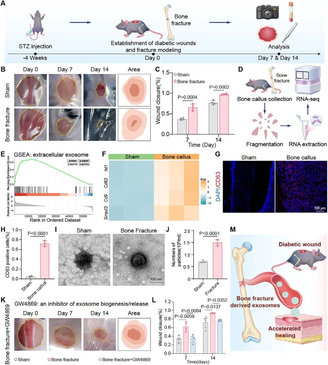
This study first established a diabetic mouse model with full-thickness skin wounds combined with femoral fracture and found that fracture accelerates wound healing. Through exosome release inhibition, small RNA sequencing of serum exosomes, and validation in callus and skin tissues, miR‑130b‑3p was identified. This miRNA targets SOCS5, promotes endothelial tube formation and Schwann cell migration, and enhances bidirectional paracrine signaling between endothelial cells and Schwann cells. For delivery of this miRNA, cholesterol-modified agomir self-assembled nanocomplexes were designed and embedded within photocrosslinkable ColMA/SF hydrogel to form a biomimetic matrix with local retention and sustained release.
03 Study Design
Streptozotocin was used to induce diabetes in mice, and a model of dorsal full-thickness wound plus femoral fracture was established. Groups with or without fracture and exosome inhibitor intervention were compared. Serum and callus exosomes were isolated for small RNA sequencing, qPCR and marker identification. The function of miR‑130b‑3p was validated in HUVECs, Schwann cells and co-culture systems, and dual-luciferase reporter assay was performed to verify targeting of SOCS5. Agomir nanocomplexes were prepared and characterized for particle size, zeta potential, nuclease stability and endocytic pathways. The formulation of ColMA/SF hydrogel was optimized, and its photocrosslinking, rheology, degradation, antioxidant capacity and release profile were evaluated. In vivo animal experiments included blank, hydrogel, free agomir and agomir-loaded hydrogel groups. Wound closure, histology, immunofluorescence, iDISCO+ tissue clearing and light-sheet three-dimensional imaging were used to assess neurovascular networks.
04 Results
Diabetic wounds exhibited accelerated healing after fracture, with enhanced re-epithelialization and collagen deposition. Circulating exosomes increased, and inhibition of exosome release abrogated the beneficial effects of fracture. miR‑130b‑3p was upregulated in serum, callus and skin and targeted SOCS5. The mimic promoted endothelial tube formation, Schwann cell migration and bidirectional paracrine signaling. Cholesterol-modified agomir self-assembled into nanocomplexes that resisted nuclease degradation, entered cells via lipid raft/caveolae-mediated endocytosis and partially escaped lysosomes. The ColMA/SF hydrogel enabled in situ gelation, sustained release, antioxidant activity and tunable degradation. A single in vivo administration of agomir hydrogel persistently elevated miR‑130b‑3p in wounds and facilitated reconstruction of vascular and nerve fiber networks. Three-dimensional imaging revealed that repair progressed from discrete foci to continuous networks, with no obvious abnormalities observed in major organs or serum biochemical parameters.
05 Outlook
This study translates bone‑skin interorgan crosstalk into a miRNA nanoformulation, demonstrating that exosomal miRNAs can act as systemic signals linking bone repair and distant tissue regeneration. Future work should clarify the cellular origin and heterogeneity of fracture-derived exosomes, dissect the target network of miR‑130b‑3p beyond SOCS5, evaluate the structural integrity and release kinetics of agomir in the wound enzymatic microenvironment, and optimize the matching between hydrogel degradation timeline and neurovascular regeneration. Robustness should be validated in models with comorbidities, across different sexes and in larger animal species, with attention to long-term material retention and systemic safety.
Source:
Advanced Science
Pub Date: 2026-07-11
DOI: 10.1002/advs.76500
Tao Shen, Hongdou Shen, Weiyue Wu, Zhuoying Jiang, Bin Liu, Yansi Xian, Rui Peng, An Yan, Yu Ben, Yong Shi, Xiang Chen, Baosheng Guo, Qing Jiang, Weijun Wang, Pingqiang Cai
We focus on custom development of functional materials in the field of nanomedicine. We tailor biofunctional nanomaterials according to your experimental requirements, regulating material structure, particle size and surface modification, with a full suite of characterization tests available. Schemes can be iteratively optimized with continuous technical support to support research projects related to targeted delivery, bioimaging, nanotheranostics and more. Feel free to share your performance specifications for customized solution evaluation.
Engineer Zhang: 17715390137
Engineer Chen: 18914047343