已传文件:photo/1794794378.png
 Biofilm formation in implant-associated infections establishes an immunosuppressive microenvironment and confers profound antibiotic tolerance. Through multi-omics analysis of clinical IAI samples, we identified lactate as a key metabolite linked to immunosuppression within the microenvironment associated with IAI biofilms. Inspired by this finding, we engineered a nanotherapeutic platform (LMVM) consisting of vacancy-engineered hollow magnetite nanoparticles co-loaded with lactate oxidase and metronidazole. By depleting lactate, LMVM remodels the immunosuppressive microenvironment and restores antibacterial immunity, highlighting lactate as an immunometabolic regulator in biofilm-associated infections. Meanwhile, lactate oxidation exacerbates local hypoxia, redirecting bacterial metabolism to activate metronidazole against tolerant subpopulations and suppressing staphyloxanthin-dependent antioxidant defense. Accordingly, this immunometabolism-guided nanotherapy achieves robust biofilm eradication and facilitates tissue repair in a mouse IAI model. Collectively, these findings establish lactate-mediated immunosuppression as a targetable feature of biofilm-associated infections and support a clinically informed material strategy that integrates immunometabolic microenvironment remodeling with the eradication of antibiotic-tolerant biofilms.

This study was published in Advanced Materials under the title “Lactate‐Depleting Nanotherapy Reprograms the Immunometabolic Microenvironment to Overcome Biofilm Tolerance in Implant‐Associated Infections”.
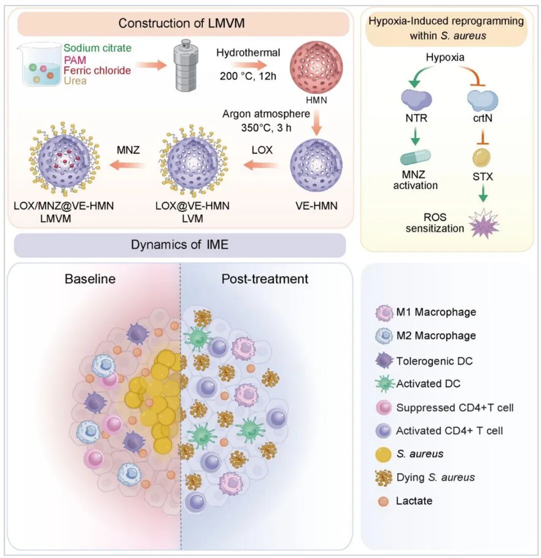
Figure 1
Schematic illustration of the fabrication and therapeutic mechanism of LMVM. LMVM is constructed by co-loading LOX and MNZ into VE-HMN. It depletes lactate to restore immune function, while exacerbating local hypoxia to enhance MNZ-mediated eradication of tolerant bacterial populations and attenuate bacterial antioxidant defense. (IME: infectious microenvironment).
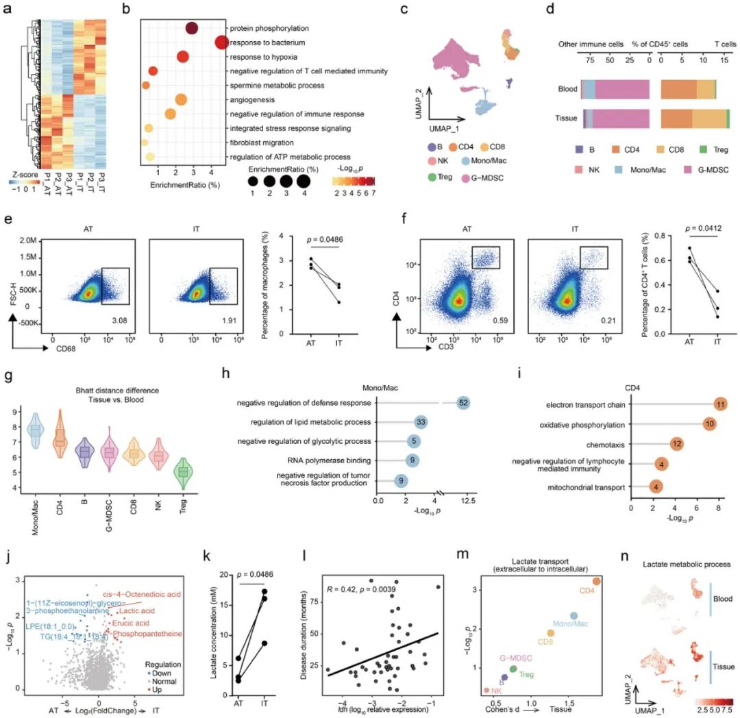
Figure 2
Multi-omics analysis of IAI. (a) Heatmap of differentially expressed genes between IT and paired AT. (b) Functional enrichment analysis of DEGs. (c) UMAP visualization of major cell types identified by scRNA-seq. (d) Frequencies of major cell types in each sample. (e,f) Flow cytometric analysis of macrophages (e) and CD4⁺ T cells (f) in AT and IT, including representative plots and quantification. (g) Box and violin plots showing the Bhattacharyya distance between tissue and blood across different cell types. (h,i) Lollipop plots illustrating upregulated biological functions in Mono/Mac (h) and CD4⁺ T cells (i). (j) Volcano plot of differentially abundant metabolites between IT and AT. (k) Lactate concentrations in IT and AT. (l) Correlation between ldh expression of Staphylococcus aureus and disease duration. Each dot represents one sample. (m) Predicted lactate transport activity in major cell types. (n) Activity of lactate metabolic processes in major cell types. Statistical significance in (e), (f), and (k) was assessed using a two-tailed paired t-test, and the correlation in (l) was evaluated by the Pearson correlation coefficient.
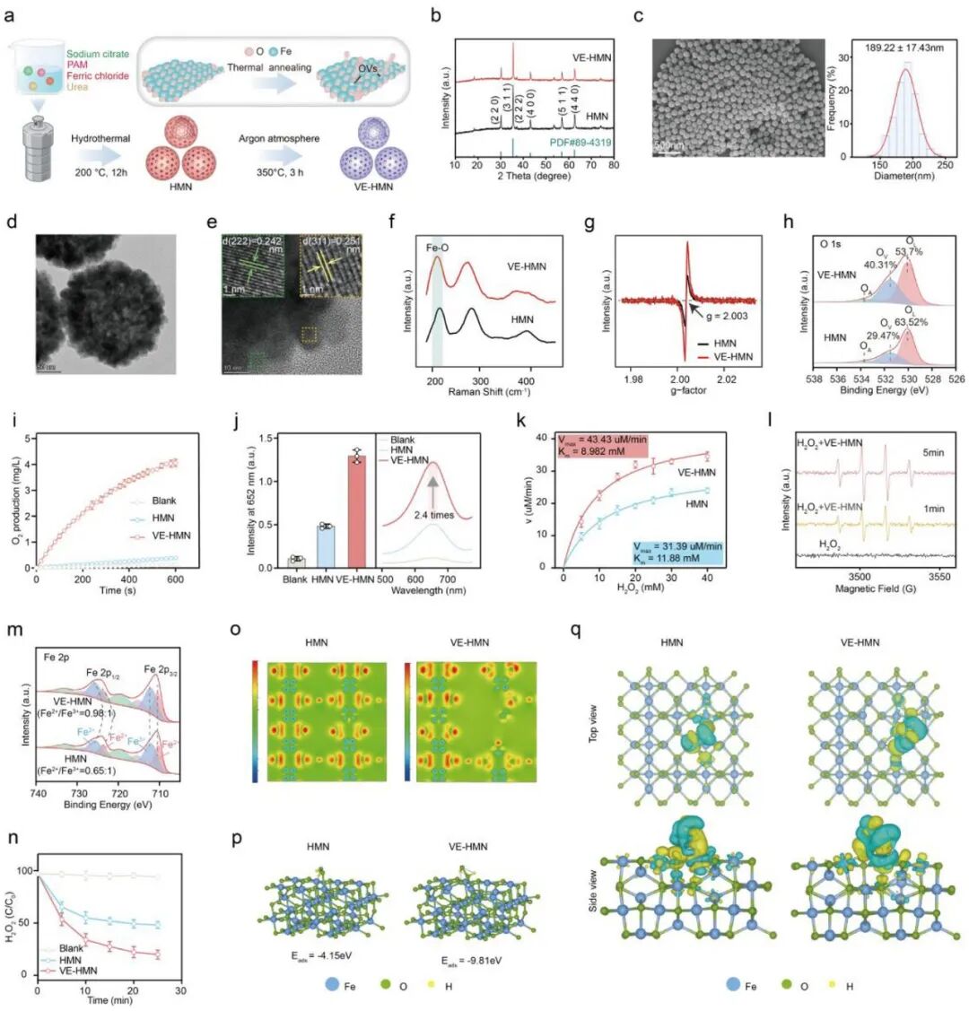
Figure 3
Synthesis, characterization and catalytic performance of VE-HMN. (a) Schematic illustration of VE-HMN synthesis. (b) XRD patterns of HMN and VE-HMN. (c) SEM image of HMN and the corresponding size distribution. (d,e) TEM (d) and HRTEM (e) images of VE-HMN. (f–h) Raman spectra (f), ESR spectra (g) and high-resolution O 1s XPS spectra (h) of HMN and VE-HMN. (i) Oxygen production from H₂O₂ catalyzed by HMN and VE-HMN. (j) Quantification and UV–vis spectra of the POD-like activity of HMN and VE-HMN. (k) Michaelis–Menten kinetics of the POD-like activity of HMN and VE-HMN using H₂O₂ as the substrate. (l) ESR spectra for the detection of ·OH signals. (m) High-resolution Fe 2p XPS spectra of HMN and VE-HMN. (n) Time-dependent H₂O₂ consumption catalyzed by HMN and VE-HMN. (o) Calculated charge density distribution of the optimized models of HMN and VE-HMN. The red and blue regions represent electron accumulation and depletion, respectively. (p) Calculated adsorption energies of H₂O₂ on HMN and VE-HMN. (q) Charge density difference of H₂O₂ adsorbed on HMN and VE-HMN. The blue and yellow regions represent electron depletion and accumulation, respectively. Data in (i), (j), (k) and (n) are presented as mean ± s.d. from three independent experiments.
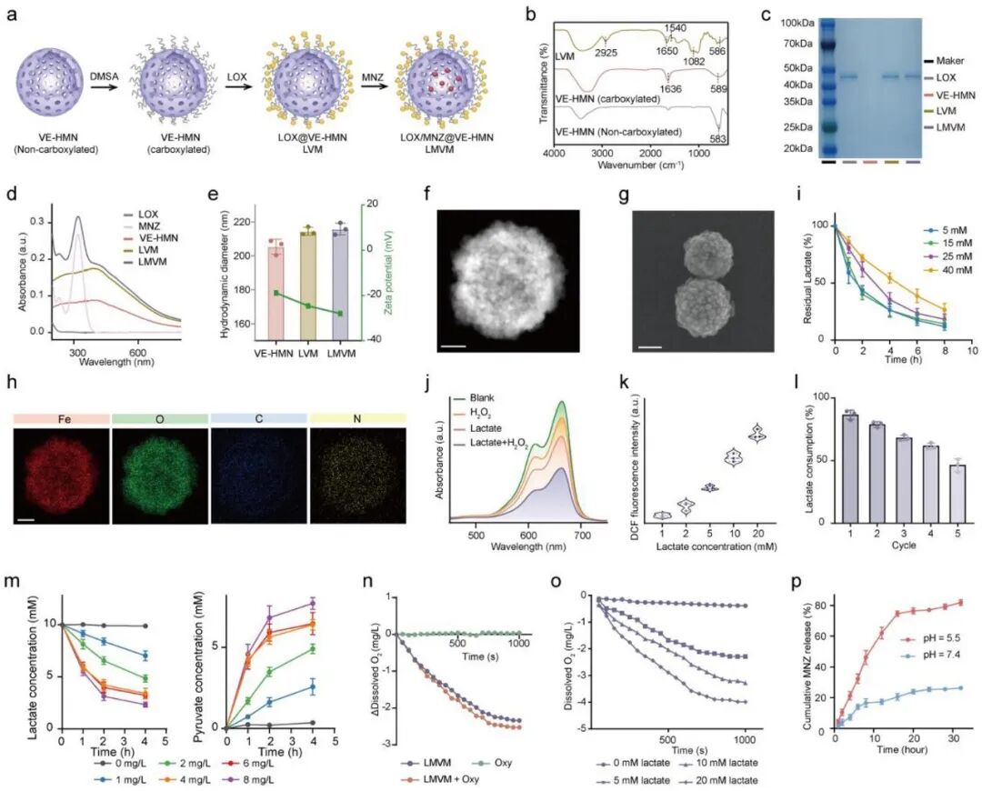
Figure 4
Fabrication, characterization and catalytic activity of LMVM. (a) Schematic illustration of the stepwise fabrication of LMVM. (b) FTIR spectra of uncarboxylated VE-HMN, carboxylated VE-HMN and LVM. (c) SDS-PAGE analysis of LOX, VE-HMN, LVM and LMVM. (d) UV–vis spectra of LOX, MNZ, VE-HMN, LVM and LMVM. (e) Hydrodynamic diameter and zeta potential distribution of VE-HMN, LVM and LMVM. (f–h) HAADF image (f), SEM image (g) and elemental mapping (h) of LMVM. Scale bars: 50 nm (f), 100 nm (g), and 50 nm (h). (i) Time-dependent lactate consumption by LMVM under different initial lactate concentrations. (j) UV–vis spectra of MB solution containing lactate, H₂O₂, or both in the LMVM catalytic system. (k) DCF fluorescence intensity in the LMVM catalytic system at different lactate concentrations. (l) Recyclable catalytic performance of LMVM, expressed as the percentage of lactate consumption in each cycle. (m) Lactate consumption (left) and pyruvate production (right) by LMVM under different dissolved oxygen conditions. (n) Dissolved oxygen profiles of LMVM with or without oxyresveratrol. (o) Oxygen consumption by LMVM at different lactate concentrations. (p) MNZ release profiles from LMVM under different pH conditions. Data in (e), (i), (l), (m) and (p) are presented as mean ± s.d. from three independent experiments.
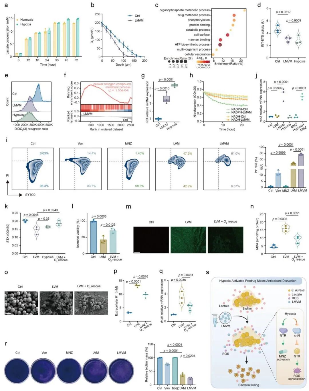
Figure 5
In vitro antibiofilm activity and mechanism of LMVM. (a) Time-dependent lactate accumulation in Staphylococcus aureus biofilm culture medium. (b) Oxygen microprofiles of biofilms with or without LMVM treatment. (c) Functional enrichment analysis of DEGs between LMVM-treated and untreated biofilms. (d,e) Electron transport system activity and bacterial membrane potential in biofilms, determined by INT reduction assay (d) and flow cytometry after DiOC₂(3) staining (e), respectively. (f) GSEA of DEGs between LMVM-treated and untreated biofilms. (g) Relative expression level of ntrA measured by RT-qPCR. (h) NTR activity in LMVM-treated and untreated biofilms evaluated by monitoring the absorbance change of nitrofurantoin in the presence of NADH or NADPH. (i) Flow cytometric analysis of live and dead cells within biofilms, showing representative plots and quantification. (j) Relative expression level of recA measured by RT-qPCR. (k) STX levels in treated biofilms. (l–q) Bacterial viability within biofilms under the indicated treatments (l), fluorescence images of ROS in biofilms stained with DCFH-DA (m), malondialdehyde content (n), SEM images of biofilms (o), potassium ion leakage (p), and relative expression level of dnaK measured by RT-qPCR (q). Scale bars, 200 µm (m) and 1 µm (o). (r) Crystal violet staining of treated biofilms, showing representative images and quantification. (s) Schematic illustration of the antibiofilm mechanism of LMVM. Data in (a), (b), (i), (l) and (r) are presented as mean ± s.d. from three independent experiments. Statistical significance in (d), (g), (i), (j), (k), (l), (n), (p), (q) and (r) was assessed using one-way analysis of variance (one-way ANOVA).
Summary
In implant-associated infections, biofilm formation creates an immunosuppressive microenvironment and confers antibiotic tolerance. Through multi-omics analysis of clinical samples, we identified lactate as a key metabolite strongly linked to immunosuppression within the biofilm-associated microenvironment. On this basis, a vacancy-engineered hollow magnetite nanoplatform co-loaded with lactate oxidase and metronidazole was designed. This platform depletes lactate to remodel the immune microenvironment and restore antibacterial immunity. Meanwhile, lactate oxidation exacerbates local hypoxia, redirects bacterial metabolism, activates metronidazole to eliminate tolerant subpopulations, and suppresses staphyloxanthin-dependent antioxidant defense, achieving biofilm eradication and tissue repair in mouse models.
This work elevates lactate from a metabolic waste product to an actionable target for biofilm-associated immunosuppression and establishes an “immunometabolism-guided” nanotherapeutic framework. In future studies, the loading dosage of lactate oxidase or the degree of hypoxia can be tuned for different infection sites to match the metabolic characteristics of bacterial strains. This strategy is extendable to other biofilm-associated infections, such as chronic osteomyelitis and catheter-related infections. The coupling between the anaerobic activation property of metronidazole and hypoxic microenvironments also offers a sequential activation concept for the design of other prodrugs. The potential impacts of prolonged hypoxia on normal tissues require further evaluation; nevertheless, this platform provides a feasible translational route for the synergy between immunometabolic modulation and antibacterial therapy.
Reference
DOI: 10.1002/adma.75214
We specialize in the custom fabrication of functional nanomedicine materials. Materials can be tailored according to experimental requirements with controllable structure, particle size and surface modification. Full characterization services are available. Research schemes can be iteratively optimized with continuous technical support. Our services support research projects including targeted delivery, bioimaging and nanotheranostics. Feel free to send your specifications for solution evaluation.
Mr. Zhang: +86 17715390137
Mr. Chen: +86 18914047343